. 2006 Sep;49(3):265-73.
doi: 10.1111/j.1365-2559.2006.02478.x.

The human renal lymphatics under normal and pathological conditions

Affiliations

The human renal lymphatics under normal and pathological conditions

Y Ishikawa et al. Histopathology. 2006 Sep.

Abstract

Aims: The renal lymphatics have not been fully documented in humans. The aim of this study was to clarify the morphology of the human renal lymphatic system under normal and pathological conditions by immunohistochemistry using anti-D2-40 antibody.

Methods and results: Normal and pathological renal tissues obtained at autopsy as well as nephrectomy specimens with renal cell carcinoma (RCC) were used. Thin sections were immunostained with antibodies against D2-40 and CD31. In normal kidney, D2-40+ lymphatics were abundant in the interstitium around the interlobar and arcuate arteries/veins but sporadic in those around the glomeruli or between the tubules in the cortex. A few lymphatics contained erythrocytes in their lumina. Lymphatics were seldom present in the medulla. In RCC cases, lymphatics were evident at the tumour margin, whereas CD31+ capillaries were abundant throughout the tumour and lymphatics were increased in the fibrous interstitium around the tumour. Lymphatic invasion by RCC cells was also detectable. D2-40+ lymphatics were evident in other pathological conditions and end-stage kidney had a denser lymphatic distribution than normal kidney.

Conclusions: Lymphatics are abundant around the arteries/veins and are also present in the renal cortex and medulla. D2-40 immunostaining is helpful for investigating the pathophysiological role of renal lymphatics.


Figures

Figure 1
Figure 1
D2-40 and von Willebrand factor immunostaining of gastric and renal tissues. a, D2-40 immunostaining of gastric tissue. D2-40 is positive in lymphatic capillaries just under the muscularis mucosae, but negative in blood vessels indicated by arrows. b, Immunohistochemistry with von Willebrand factor in a serial section of that seen in (a). Blood vessels indicated by arrows in (a) are positive, but lymphatic capillaries indicated by arrows in this figure are negative. c, D2-40 immunostaining of the kidney. Capillaries around the arteries in the cortex are positive for D2-40 antibody, but the endothelia of the arteries and glomeruli are negative. d, Negative control section of (c) by omitting the primary antibody and replacing it with normal mouse immunoglobulin. The figure is a serial section of that seen in (c), which demonstrates negative staining.
Figure 2
Figure 2
Lymphatics in the normal kidney (D2-40 immunostaining). b, Lymphatic capillaries are evident in the interstitium around the glomerulus. c, Lymphatics exhibiting a slit-like structure are distributed around the interlobular artery and vein in the cortex. c, In the interstitium around a dilated interlobular vein, many lymphatic capillaries are evident. A few lymphatic capillaries are present just beneath the venous endothelium. d, A lymphatic capillary is recognizable in the centre of the figure showing a normal medulla.
Figure 3
Figure 3
Large blood vessel-related lymphatics in the normal kidney. a, Lymphatic vessels positive for D2-40 are abundantly distributed in the interstitium around the interlobar artery. b, Many lymphatics are shown around the interlobar veins. Longitudinal aspect of a lymphatic vessel is recognized in the interstitium adjacent to a central vein. c, Elastica van Gieson staining demonstrates a vessel (arrowheads) containing erythrocytes in the lumen and running alongside the interlobar vein. d, Figure showing the same area as (c) in a serial section. A vessel (arrowheads) containing erythrocytes found in (c) is positive for D2-40, which indicates that it is a lymphatic vessel. e, The arcuate vein in the centre of the figure shows positivity for CD31. Capillaries adjacent to the vein are also immunopositive for CD31 (arrows). f, Figure showing the same area as (d) in a serial section immunostained with D2-40. Note two capillaries (arrows) positive for D2-40, which are also positive for CD31, as shown in (e).
Figure 4
Figure 4
Lymphatic distribution in the normal kidney in relation to anatomical structures. This figure shows the number of lymphatics in 20 high-power fields (× 200) in 10 cases with normal structures. The values are 12.0 ± 7.9 in the interstitium between the tubules in the cortex (Cortex), 7.4 ± 3.6 in the interstitium around the glomeruli (Glomerulus), 0.8 ± 1.7 in the interstitium of the medulla (Medulla), 36.0 ± 9.4 in the interstitium around the interlobular arteries/veins (Interlobular) and 78.3 ± 24.5 in the interstitium around the interlobar arteries/veins (Interlobar). The lymphatics are abundant in the interstitium around the arteries/veins. Hardly any lymphatics are evident in the medulla. Statistical analyses (Student's t-test) between all location groups revealed a significant difference of P < 0.0001 except for comparison between ‘Cortex’ and ‘Interlobar’, which was P = 0.0236.
Figure 5
Figure 5
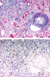
Lymphatics in end-stage kidney. a, Azan–Mallory staining of an end-stage kidney demonstrates severe atrophy of the cortex. The interlobular artery exhibits fibrous thickening of its wall. b, Figure showing the same area of a serial section of (a). In the interstitium of the cortex, lymphatic capillaries positive for D2-40 are abundantly distributed, but artery-related lymphatics are scarce.
Figure 6
Figure 6
Comparison of lymphatic distribution in the normal cortex with that in end-stage kidney, fibrous cortex around renal cell carcinoma (RCC) and the intra-RCC area.The number of lymphatics in 20 high-power fields (× 200) is shown for four categorized locations. The values are 18.6 ± 4.6 in the normal cortex (n = 10), 27.6 ± 2.6 in the cortex of end-stage kidney (n = 3), 207.7 ± 105.9 in fibrous cortex around RCC (peri-RCC cortex; n = 10) and 0.8 ± 1.5 in the stroma of RCC (intra-RCC; n = 10). The number of lymphatics is significantly higher in the cortex of end-stage kidney than in the normal kidney. In RCC cases, the peri-RCC cortex shows abundant lymphatic distribution that is significantly more extensive than that of normal kidney, but lymphatic vessels are seldom detected in the area of intra-RCC. *The value for the intra-RCC area is significantly lower (P < 0.0001) than those for the other three locations.
Figure 7
Figure 7
Immunohistochemistry of kidney with renal cell carcinoma (RCC). a, The central area of RCC has abundant blood vessels positive for CD31. b, Figure showing the same area in a serial section of (a). D2-40 immunostaining reveals a negative result. c, At the tumour margin, two lymphatics positive for D2-40 are invaded by tumour cells (arrows). d, In the fibrous interstitium around the tumour, lymphatics positive for D2-40 are abundantly distributed. Arrowheads indicate the boundary between the tumour and the tumour-free cortex.

References

    1. Rohn DA, Stewart RH, Elk JR, Laine GA, Drake RE. Renal lymphatic function following venous pressure elevation. Lymphology. 1996;29:67–75. - PubMed
    1. Holmes MJ, O'Morchoe PJ, O'Morchoe CCC. Morphology of the intrarenal lymphatic system; capsular and hilar communications. Am. J. Anat. 1977;149:333–352. - PubMed
    1. Hogg CM, Reid O, Scothorne RJ. Studies on hemolymph nodes. III. Renal lymph as a major source of erythrocytes in the renal hemolymph node of the rat. J. Anat. 1982;135:291–299. - PMC - PubMed
    1. Cuttino JT, Jr, Jennette JC, Clark RL, Kwock L. Renal medullary lymphatics: microradiographic, light, and electron microscopic studies in pigs. Lymphology. 1985;18:24–30. - PubMed
    1. Cuttino JT, Jr, Clark RL, Jennette JC. Microradiographic demonstration of human intrarenal microlymphatic pathways. Urol. Radiol. 1989;11:83–87. - PubMed

Publication types

Substances