. 2006 Sep 15;362(2):173-83.
doi: 10.1016/j.jmb.2006.07.046. Epub 2006 Jul 28.

B29 gene silencing in pituitary cells is regulated by its 3' enhancer

Affiliations

B29 gene silencing in pituitary cells is regulated by its 3' enhancer

Cindy S Malone et al. J Mol Biol. .

Abstract

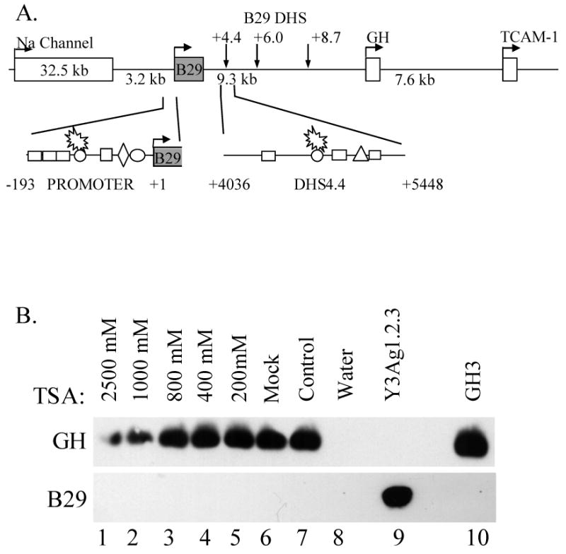
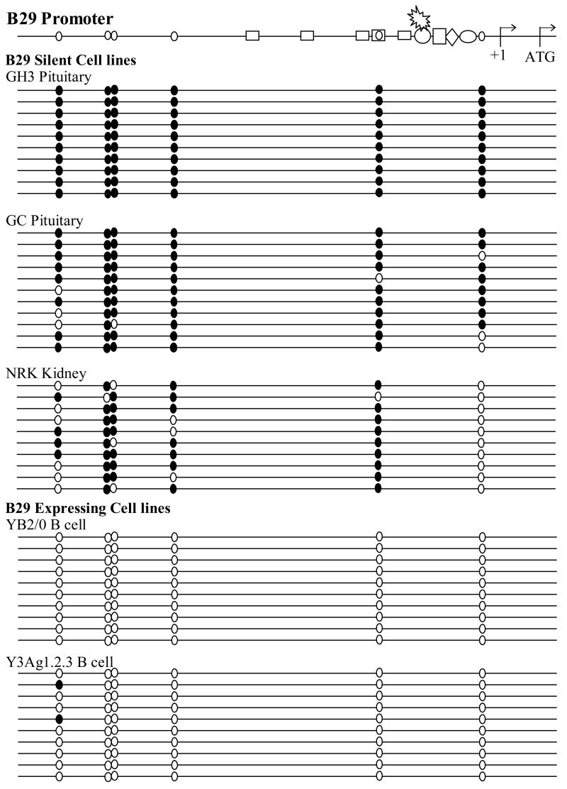
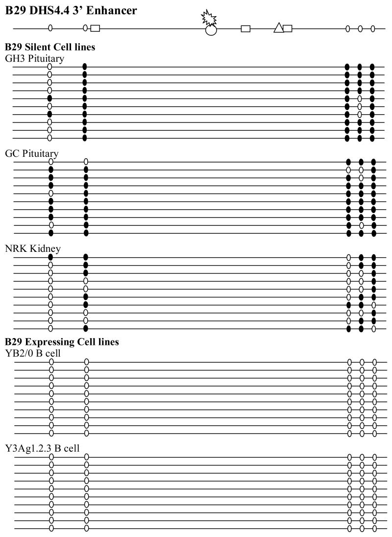
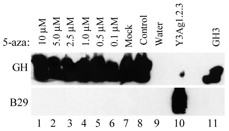
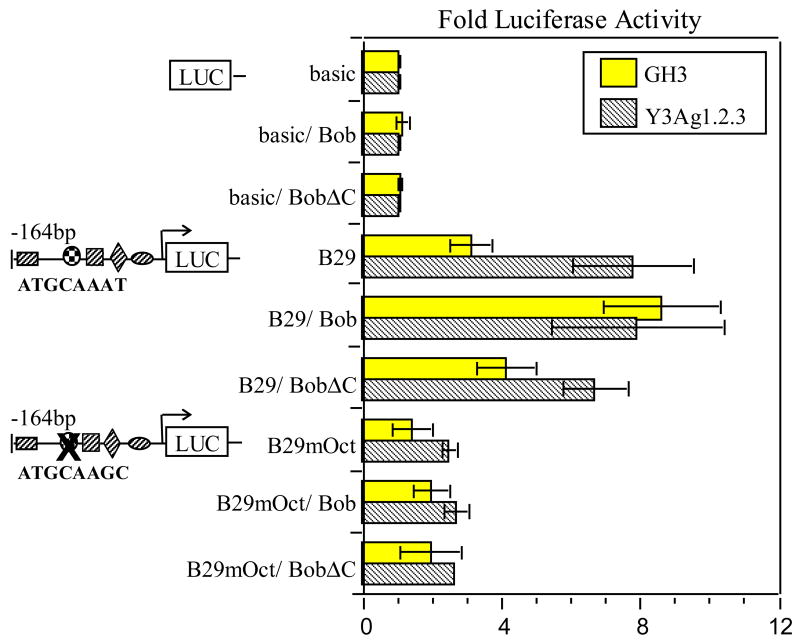
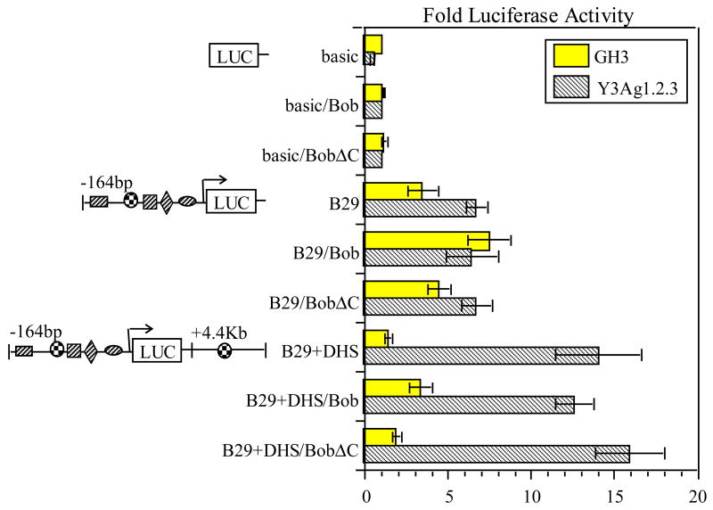
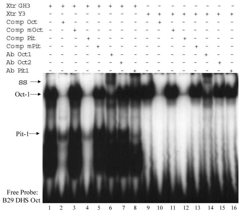
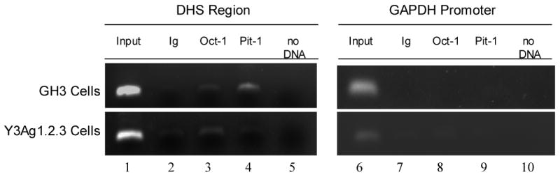
B cell-specific B29 (Igbeta, CD79b) genes in rat, mouse, and human are situated between the 5' growth hormone (GH) locus control region and the 3' GH gene cluster. The entire GH genomic region is DNase 1 hypersensitive in GH-expressing pituitary cells, which predicts an "open" chromatin configuration, and yet B29 is not expressed. The B29 promoter and enhancers exhibit histone deacetylation in pituitary cells, but histone deacetylase inhibition failed to activate B29 expression. The B29 promoter and a 3' enhancer showed local dense DNA methylation in both pituitary and non-lymphoid cells consistent with gene silencing. However, DNA methyltransferase inhibition did not activate B29 expression either. B29 promoter constructs were minimally activated in transfected pituitary cells. Co-transfection of the B cell-specific octamer transcriptional co-activator Bob1 with the B29 promoter construct resulted in high level promoter activity in pituitary cells comparable to B29 promoter activity in transfected B cells. Unexpectedly, inclusion of the B29 3' enhancer in B29 promoter constructs strongly inhibited B29 transcriptional activity even when pituitary cells were co-transfected with Bob1. Both Oct-1 and Pit-1 bind the B29 3' enhancer in in vitro electrophoretic mobility shift assay and in in vivo chromatin immunoprecipitation analyses. These data indicate that the GH locus-embedded, tissue-specific B29 gene is silenced in GH-expressing pituitary cells by epigenetic mechanisms, the lack of a B cell-specific transcription factor, and likely by the B29 3' enhancer acting as a powerful silencer in a context and tissue-specific manner.


Figures

Figure 1
Figure 1. HDAC inhibitor TSA does not reactivate the B29 gene in pituitary cells
A. Schematic diagram of the rat B29 and growth hormone (GH) genomic locus. The B29 promoter, coding sequence and 3′ enhancers are located between the muscle-specific sodium channel gene and the pituitary-specific GH gene. B29 promoter and B29 DHS4.4 3′ enhancer transcription factor binding sites are shown. Rectangle; EBF, circle; octamer, spikey oval; Bob1, square; ETS, diamond; Sp1, oval: Ikaros, triangle; NF-kB. Cartoon drawn to scale. Numbers indicate distances from the major start of B29 transcription (+1) B. Southern blots of GH and B29 RT-PCR gels were used to detect gene expression. GH3 pituitary cells were treated with TSA in increasing doses to the maximum tolerated dosage for three days before RNA was prepared, Lanes 1 – 5. Lane 6: mock TSA, lane 7: no treatment, lane 8: water, lane 9: Y3Ag1.2.3 myeloma cell RNA, lane 10: GH3 pituitary cell RNA.
Figure 2
Figure 2. Nearly complete DNA methylation of the B29 promoter in silent pituitary cells
Genomic DNA from pituitary, kidney and myeloma cell lines was subjected to sodium bisulfite conversion, PCR amplification of a 400 bp fragment, subcloning and cycle sequencing. Unmethylated CpG sites are indicated by small vertical open ovals and methylated CpG sites are indicated by small closed ovals. At least ten clones from each cell line as indicated were bisulfite sequenced to obtain a representative sampling of DNA methylation patterns in each setting. Transcription factor binding sites are as indicated: rectangle; EBF, circle; Octamer, triangle; ETS, diamond; Sp1, oval; Ikaros. +1 indicates the major transcription start site. Cartoon drawn to scale.
Figure 3
Figure 3. Dense DNA methylation of the B29 DHS4.4 3′ enhancer in pituitary cells
Genomic DNA from pituitary, kidney and myeloma cells was subjected to sodium bisulfite conversion, PCR amplification of a 400 bp fragment, subcloning and cycle sequencing. Unmethylated CpG sites are indicated by small vertical open ovals and methylated CpG sites are indicated by small closed ovals. At least ten clones from each cell line as indicated were bisulfite sequenced to obtain a representative sampling of methylation patterns in each setting. Transcription factor binding sites are by homology only (except Octamer) and are as indicated: rectangle; EBF, circle; Octamer, triangle; NF-κB. Cartoon drawn to scale.
Figure 4
Figure 4. Methylation inhibitor 5-aza does not reactivate the B29 gene in pituitary cells
Southern blots of GH and B29 RT-PCR gels were used to detect gene expression. GH3 pituitary cells were treated with 5-aza in increasing doses to the maximum tolerated dosage for three days before RNA was prepared, Lanes 1 – 6. Lane 7: mock 5-aza, lane 8: no treatment, lane 9: water, lane 10: Y3Ag1.2.3 myeloma cell RNA, lane 11: GH3 pituitary cell RNA.
Figure 5
Figure 5. B29 promoter expression is enhanced by co-expression of Bob1 in pituitary cells
Transient transfections of pGL3 B29 promoter constructs with and without co-transfection of Bob1 were conducted in B29-silent GH3 pituitary cells (shaded bar) and B29-expressing Y3Ag1.2.3 myeloma cells (striped bar). The activity of each construct is expressed as the fold activation over the promoterless pGL3 basic firefly luciferase construct. A total of 10 μg Bob1 expression construct (Bob) or 10 μg transactivation-negative Bob1 C-terminal deletion mutant construct (BobΔC) was added to transient transfections of the pGL3 basic firefly luciferase construct (basic), the pGL3 B29 promoter construct (B29), and the pGL3 B29 promoter with a mutated Octamer motif construct (B29mOct). pGL3 firefly luciferase activities are pRL SV40 renilla luciferase normalized and are the average ±SD of at least three independent transfections using at least two preparations of DNA.
Figure 6
Figure 6. Control of B29 expression by a context-dependent B29 DHS4.4 3′ enhancer molecular toggle
Transient transfections of pGL3 B29 promoter and DHS4.4 3′ enhancer constructs with and without co-transfection of Bob1 in B29-silent GH3 pituitary cells (shaded bar) and B29-expressing Y3Ag1.2.3 myeloma cells (striped bar). The activity of each construct is expressed as the fold activation over the promoterless pGL3 basic firefly luciferase construct. A total of 10 μg Bob1 expression construct (Bob) or 10 μg transactivation-negative Bob1 C-terminal deletion mutant construct (BobΔC) was added to transient transfections of the pGL3 basic firefly luciferase construct (basic), the pGL3 B29 promoter construct (B29), and the pGL3 B29 promoter and DHS 4.4 construct (B29+DHS). pGL3 firefly luciferase activities are pRL SV40 renilla luciferase normalized and are the average ±SD of at least three independent transfections using at least two preparations of DNA.
Figure 7
Figure 7. B29 DHS4.4 3′ enhancer binds Oct-1 and Pit-1 by EMSA
Double-stranded oligonucleotides corresponding to the B29 DHS4.4 3′ enhancer Octamer site (B29 DHS Oct) were end-labeled and used in EMSA. The B29 DHS Oct probe was incubated with 20 μg GH3 pituitary cell line nuclear extract (lanes 1-8) and 20 μg Y3Ag1.2.3 B cell line nuclear extract (lanes 9-16). Reactions were co-incubated in the presence of: 500-fold molar excess unlabeled B29 Octamer motif (Comp Oct, lanes 2 and 10); 500-fold molar excess unlabeled B29 mutant Octamer motif (Comp mOct, lanes 3 and 11); 500-fold molar excess unlabeled Pit-1 consensus motif (Comp Pit, lanes 4 and 12); 500-fold molar excess unlabeled Pit-1 mutant consensus motif (Comp mPit, lanes 5 and 13); 0.5 μg anti-Oct-1 antibody (Ab Oct1, lanes 6 and 14); 2 μg anti-Oct-2 antibody (Ab Oct2, lanes 7 and 15); and 1 μg anti-Pit-1 antibody (Ab Pit1, lanes 8 and 16). Specifically formed complexes are labeled “Oct-1” and “Pit-1” and denoted by arrows. Antibody supershifted complexes are denoted by “SS” and an arrow. “Free Probe” denotes uncomplexed endlabeled B29 DHS Oct probe. Results are representative of at least three independent experiments.
Figure 8
Figure 8. B29 DHS4.4 3′ enhancer binds Oct-1 and Pit-1 by ChIP
Chromatin was purified from GH3 pituitary cells (top panel) and Y3Ag1.2.3 B cells (bottom panel) and used in ChIP analyses. Chromatin was immunoprecipitated using normal rabbit immune sera (Ig, lanes 2 and 7), anti-Oct-1 antibody (Oct-1, lanes 3 and 8), and anti-Pit-1 antibody (Pit-1, lanes 4 and 9). The presence of immunoprecipitated chromatin was determined for the B29 DHS4.4 3′ enhancer region (DHS region, lanes 1-5) and the GAPDH promoter negative control (GAPDH promoter, lanes 6-10) by PCR using region-specific primers. Lanes 1 and 6 are positive control PCR reactions performed using the purified chromatin before immunoprecipitation (Input) and lanes 5 and 10 are negative control PCR reactions performed without the addition of any chromatin (no DNA).

Similar articles

Cited by

References

    1. Benschop RJ, Cambier JC. B cell development: signal transduction by antigen receptors and their surrogates. Curr Opin Immunol. 1999;11:143–51. - PubMed
    1. Hermanson GG, Eisenberg D, Kincade PW, Wall R. B29: a member of the immunoglobulin gene superfamily exclusively expressed on beta-lineage cells. Proc Natl Acad Sci U S A. 1988;85:6890–4. - PMC - PubMed
    1. Wang H, Diamond RA, Rothenberg EV. Cross-lineage expression of Ig-beta (B29) in thymocytes: positive and negative gene regulation to establish T cell identity. Proc Natl Acad Sci U S A. 1998;95:6831–6. - PMC - PubMed
    1. Omori SA, Wall R. Multiple motifs regulate the B-cell-specific promoter of the B29 gene. Proc Natl Acad Sci U S A. 1993;90:11723–7. - PMC - PubMed
    1. Thompson AA, Wood WJ, Jr, Gilly MJ, Damore MA, Omori SA, Wall R. The promoter and 5′ flanking sequences controlling human B29 gene expression. Blood. 1996;87:666–73. - PubMed

Publication types

MeSH terms

LinkOut - more resources