

Antigenic and cellular localisation analysis of the severe acute respiratory syndrome coronavirus nucleocapsid protein using monoclonal antibodies
- PMID: 16920216
- PMCID: PMC7114340
- DOI: 10.1016/j.virusres.2006.07.005
Antigenic and cellular localisation analysis of the severe acute respiratory syndrome coronavirus nucleocapsid protein using monoclonal antibodies
Abstract
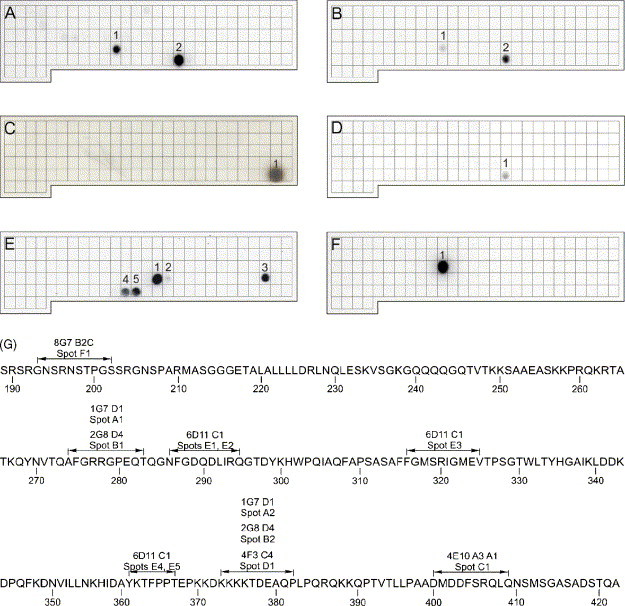
A member of the family of coronaviruses has previously been identified as the cause of the severe acute respiratory syndrome (SARS). In this study, several monoclonal antibodies against the nucleocapsid protein have been generated to examine distribution of the nucleocapsid in virus-infected cells and to study antigenic regions of the protein. Confocal microscopic analysis identified nucleocapsids packaged in vesicles in the perinuclear area indicating viral synthesis at the endoplasmic reticulum and Golgi apparatus. The monoclonal antibodies bound to the central and carboxyterminal half of the nucleocapsid protein indicating prominent exposure and immunogenicity of this part of the protein. Antibodies recognised both linear and conformational epitopes. Predictions of antigenicity using mathematical modelling based on hydrophobicity analysis of SARS nucleoprotein could not be confirmed fully. Antibody binding to discontinuous peptides provides evidence that amino acids 274-283 and 373-382 assemble to a structural unit particularly rich in basic amino acids. In addition, amino acids 286-295, 316-325 and 361-367 that represent the epitope recognised by monoclonal antibody 6D11C1 converge indicating a well-structured C-terminal region of the SARS virus nucleocapsid protein and functional relationship of the peptide regions involved. Alternatively, dimerisation of the nucleocapsid protein may result in juxtaposition of the amino acid sequences 316-325 and 361-367 on one nucleoprotein molecule to amino acid 286-295 on the second peptide. The monoclonal antibodies will be available to assess antigenicity and immunological variabilities between different SARS CoV strains.
Figures
Similar articles
-
The SARS-CoV nucleocapsid protein: a protein with multifarious activities.Infect Genet Evol. 2008 Jul;8(4):397-405. doi: 10.1016/j.meegid.2007.07.004. Epub 2007 Jul 20. Infect Genet Evol. 2008. PMID: 17881296 Free PMC article. Review.
-
Biochemical and immunological studies of nucleocapsid proteins of severe acute respiratory syndrome and 229E human coronaviruses.Proteomics. 2005 Mar;5(4):925-37. doi: 10.1002/pmic.200401204. Proteomics. 2005. PMID: 15759315 Free PMC article.
-
Characterization and epitope mapping of monoclonal antibodies to the nucleocapsid protein of severe acute respiratory syndrome coronavirus.Jpn J Vet Res. 2008 Feb;55(4):115-27. Jpn J Vet Res. 2008. PMID: 18380153
-
Molecular and biological characterization of human monoclonal antibodies binding to the spike and nucleocapsid proteins of severe acute respiratory syndrome coronavirus.J Virol. 2005 Feb;79(3):1635-44. doi: 10.1128/JVI.79.3.1635-1644.2005. J Virol. 2005. PMID: 15650189 Free PMC article.
-
Mapping of antigenic sites on the nucleocapsid protein of the severe acute respiratory syndrome coronavirus.J Clin Microbiol. 2004 Nov;42(11):5309-14. doi: 10.1128/JCM.42.11.5309-5314.2004. J Clin Microbiol. 2004. PMID: 15528730 Free PMC article.
Cited by
-
High protection and transmission-blocking immunity elicited by single-cycle SARS-CoV-2 vaccine in hamsters.NPJ Vaccines. 2024 Oct 30;9(1):206. doi: 10.1038/s41541-024-00992-z. NPJ Vaccines. 2024. PMID: 39472701 Free PMC article.
-
The SARS-CoV nucleocapsid protein: a protein with multifarious activities.Infect Genet Evol. 2008 Jul;8(4):397-405. doi: 10.1016/j.meegid.2007.07.004. Epub 2007 Jul 20. Infect Genet Evol. 2008. PMID: 17881296 Free PMC article. Review.
-
Detailed epitope mapping of SARS-CoV-2 nucleoprotein reveals specific immunoresponse in cats and dogs housed with COVID-19 patients.Res Vet Sci. 2022 Mar;143:81-87. doi: 10.1016/j.rvsc.2021.12.020. Epub 2022 Jan 4. Res Vet Sci. 2022. PMID: 34998208 Free PMC article.
-
The Ebola Virus Nucleoprotein Recruits the Nuclear RNA Export Factor NXF1 into Inclusion Bodies to Facilitate Viral Protein Expression.Cells. 2020 Jan 11;9(1):187. doi: 10.3390/cells9010187. Cells. 2020. PMID: 31940815 Free PMC article.
-
Characterization of SARS-CoV-2 N protein reveals multiple functional consequences of the C-terminal domain.bioRxiv [Preprint]. 2020 Nov 30:2020.11.30.404905. doi: 10.1101/2020.11.30.404905. bioRxiv. 2020. Update in: iScience. 2021 Jun 25;24(6):102681. doi: 10.1016/j.isci.2021.102681. PMID: 33269347 Free PMC article. Updated. Preprint.
References
-
- Davies H.A., Dourmashkin R.R., Macnaughton M.R. Ribonucleoprotein of avian infectious bronchitis virus. J. Gen. Virol. 1981;53(Pt 1):67–74. - PubMed
MeSH terms
Substances
LinkOut - more resources
Full Text Sources
Other Literature Sources
Molecular Biology Databases
Miscellaneous
