

Therapeutic implications of autoimmune vitiligo T cells
- PMID: 16920575
- PMCID: PMC3462656
- DOI: 10.1016/j.autrev.2006.03.012
Therapeutic implications of autoimmune vitiligo T cells
Abstract
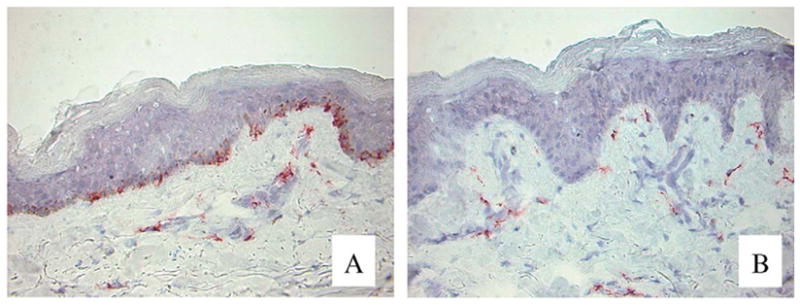
Vitiligo is an autoimmune disease presenting with progressive loss of skin pigmentation. The disease strikes 1% of the world population, generally during teenage years. The progressive loss of melanocytes from depigmenting vitiligo skin is accompanied by cellular infiltrates containing both CD4+ and CD8+ T lymphocytes. Infiltrating cytotoxic T cells with high affinity T cell receptors have likely escaped clonal deletion in the thymus, allowing such T cells to enter the circulation. Through the expression of CLA, these T cells home to the skin where they express type 1-cytokine profiles and mediate melanocyte apoptosis via the granzyme/perforin pathway. T cells found juxtapositionally apposed to remaining melanocytes can be isolated from the skin. Vitiligo T cells have demonstrated reactivity to antigens previously recognized as target antigens for T cells infiltrating melanoma tumors. In a comparison to existing melanoma-derived T cells, vitiligo T cells displayed superior reactivity towards melanoma cells. It is thought that genes encoding the TCRs expressed by vitiligo skin infiltrating T cells can be cloned and expressed in melanoma T cells, thereby generating a pool of circulating T cells with high affinity for their targets that can re-direct the immune response towards the tumor.
Figures

References
-
- Le Poole IC, van den Wijngaard RMJGJ, Westerhof W, Dutrieux RP, Das PK. Presence or absence of melanocytes in vitiligo lesions: an immunohistochemical investigation. J Invest Dermatol. 1993;100(6):816–33. - PubMed
-
- Liu JB, Li M, Yang S, Gui JP, Wanf HY, Du WH, et al. Clinical profiles of vitiligo in China: an analysis of 3742 patients. Clin Exp Dermatol. 2005;30(4):327–31. - PubMed
-
- Dogra S, Parsad D, Handas S, Kanwar AJ. Late onset vitiligo: a study of 182 patients. Int J Dermatol. 2005;44(3):193–6. - PubMed
-
- Halder RM, Nootheti PK. Ethnic skin disorders overview. J Am Acad Dermatol. 2003;48(S6):S143–8. - PubMed
-
- Chaturvedi SK, Singh G, Gupta N. Stigma experience in skin disorders: an Indian perspective. Dermatol Clin. 2005;23 (4):635–42. - PubMed
Publication types
MeSH terms
Substances
Grants and funding
LinkOut - more resources
Full Text Sources
Other Literature Sources
Medical
Research Materials
