Comparative Study
. 2006;34(15):4245-57.
doi: 10.1093/nar/gkl521. Epub 2006 Aug 18.

Mycobacterial transcriptional signals: requirements for recognition by RNA polymerase and optimal transcriptional activity

Affiliations
Comparative Study

Mycobacterial transcriptional signals: requirements for recognition by RNA polymerase and optimal transcriptional activity

Nisheeth Agarwal et al. Nucleic Acids Res. 2006.

Abstract

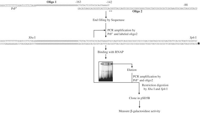
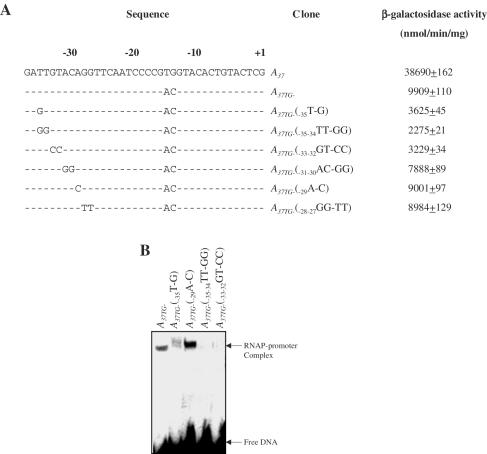
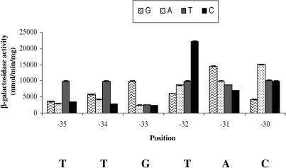
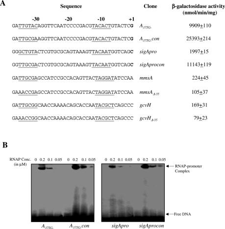
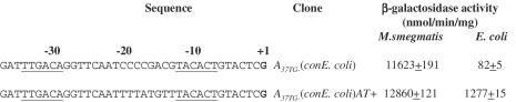
Majority of the promoter elements of mycobacteria do not function well in other eubacterial systems and analysis of their sequences has established the presence of only single conserved sequence located at the -10 position. Additional sequences for the appropriate functioning of these promoters have been proposed but not characterized, probably due to the absence of sufficient number of strong mycobacterial promoters. In the current study, we have isolated functional promoter-like sequences of mycobacteria from the pool of random DNA sequences. Based on the promoter activity in Mycobacterium smegmatis and score assigned by neural network promoter prediction program, we selected one of these promoter sequences, namely A37 for characterization in order to understand the structure of housekeeping promoters of mycobacteria. A37-RNAP complexes were subjected to DNase I footprinting and subsequent mutagenesis. Our results demonstrate that in addition to -10 sequences, DNA sequence at -35 site can also influence the activity of mycobacterial promoters by modulating the promoter recognition by RNA polymerase and subsequent formation of open complex. We also provide evidence that despite exhibiting similarities in -10 and -35 sequences, promoter regions of mycobacteria and Escherichia coli differ from each other due to differences in their requirement of spacer sequences between the two positions.


Figures

Figure 1
Figure 1
Generation of promoter fragments with random sequences by in vitro selection. Oligonucleotides were annealed and extended with Sequenase DNA polymerase to form a library of double-stranded DNA fragments carrying 29 bp long random sequence, flanked by constant sequences, at its 5′ and 3′ ends. The flanking sequence at the 5′ end comprised of a partial TER3 terminator sequence of pSD5B and the flanking sequence at the 3′ end carried the sequence of coding strand of sigA gene from position −142 to −163 (with respect to translational start codon). The oligo2 contained the mutant sequence (positions of mutations are marked by small asterisks) of template strand of sigA gene from position −88 to −163 (with respect to translational start codon). These oligonucleotides were designed in such a way that the resulting double-stranded DNA fragments contained an XbaI site upstream to random sequence and an SphI site downstream to it. The end-labeled DNA fragments were obtained by subjecting the double-stranded DNA fragment to PCR amplification by using primer PrF′ and labeled primer oligo2. Primers, PrF′ and oligo2, were designed in such a way that the amplicon carried sufficient bases (76 bases) downstream to the random sequence to ensure that the selected promoter sequences have sufficient bases downstream to the TSP. These end-labeled DNA fragments were subjected to EMSA by allowing them to form the complex with RNAP of M.smegmatis. The DNA fragments were subsequently eluted from the gel and re-amplified by PrF′ and oligo2. PCR amplicon was subsequently used for either its cloning into the pSD5B (after digestion by XbaI and SphI) or as a template for second round of PCR amplification using primer PrF′ and labeled primer oligo2. β-Galactosidase activity of the promoter clones was determined, as described in Materials and Methods. Bold star represents the labeled end of promoter DNA fragment.
Figure 2
Figure 2
Analysis of unique promoter clones. The open box represents the position of 29 bp long unique sequence in each promoter clone, flanked by constant sequences, at its 5′ and 3′ ends, as described in Figure 1. Promoter activity was measured as β-galactosidase specific activity. The values represent mean of three separate assays.
Figure 3
Figure 3
Promoter prediction by the NNPP program. Each of the DNA sequences containing 29 bp long unique sequence, flanked by 15 bp long constant sequences, at its 5′ and 3′ ends was used for the promoter prediction by NNPP program by using minimum promoter score >0.25. +1 indicates the putative TSP, identified by the NNPP program. The values represent the score assigned to each DNA sequence.
Figure 4
Figure 4
Analysis of A37 promoter. (A) Schematic illustration of pSD5B-A37 promoter clone. Relevant restriction sites, positions of various primers (PrF, PrR and PrlacZ) and antibiotic resistance marker (kn) are shown. TER1, TER2 and TER3 represent the transcriptional terminators, as described previously (24,25). (B) Determination of TSP of A37. Primer extension reaction was carried out with end-labeled primer PrlacZ and RNA isolated from the M.smegmatis cells harboring pSD5B-A37 vector. The reactions were analyzed on urea–6% polyacrylamide gels. Sequencing reactions were also performed with PrlacZ and run alongside the corresponding primer extension reactions (lane P). Arrow represents position of TSP, which is shown as a bold letter in the sequence. (C) Sequence analysis of A37 promoter containing region of pSD5B-A37. +1 represents TSP of A37. Sequences written in bold indicate position of −10 sequence of A37. Horizontal arrows represent the sequences of PrF, PrR and PrlacZ primers, respectively. Sequence written in italics represents the transcriptional terminator, TER3. Positions of restriction sites flanking the promoter region are shown. (D) DNase I protection assay using RNAP bound to the A37 promoter. The gel electrophoretic pattern and the corresponding densitometric scan of the coding strand are shown. Arrows indicate the positions of DNase I hyperactive sites due to the binding of RNAP. The numbers indicate the positions of respective bases. The solid vertical bar indicates the positions on the promoter region that are completely protected; the interrupted bar represents the sequence that shows partial protection. G+A represents sequencing ladder by the Maxam–Gilbert method; ‘+’ and ‘−’ represent DNase I reactions with and without RNAP, respectively. Densitometric scanning was performed by using NIH Image program.
Figure 5
Figure 5
Base substitutions at −35 region and its influence on promoter functions. (A) Effects of base substitutions on the activity of A37TG- promoter. Promoter activity was measured as β-galactosidase specific activity. The cell lysate of pSD5B-transformed M.smegmatis (exhibiting an activity of 12 ± 5 nmol/min/mg; data not shown) was used as a negative control. The values represent mean of three separate assays. ‘-’ represents similar bases at the corresponding position. The numbers above promoter sequence indicate the positions of respective bases. (B) EMSA with wild-type and mutant A37TG- promoter fragments. Binding reactions containing 1.5 nM of radiolabeled promoter DNA fragment and 0.1 μM RNA polymerase were carried out at 37°C for 10 min. The reactions were terminated by addition of 10 μg/ml heparin for 2 min. After incubation, samples were loaded on 4% polyacrylamide (30:1 acrylamide/Bis) gel.
Figure 6
Figure 6
Effects of single-base substitutions on the activity of A37TG- promoter in M.smegmatis. All three possible substitutions were carried out at each position from −35 to −30. The wild-type sequence at each position is shown below the figure. Promoter activity was measured as β-galactosidase specific activity. The cell lysate of pSD5B-transformed M.smegmatis (exhibiting an activity of 12 ± 5 nmol/min/mg; data not shown) was used as a negative control. The values represent mean of three separate assays.
Figure 7
Figure 7
Functional analysis of promoter derivatives with altered sequence in the −35 region. (A) Comparative analysis of the activities of wild-type and mutant derivatives of mycobacterial promoters, respectively. sigA promoter fragment contains the promoter sequence from position −143 to +42 with respect to its TSP and both the mmsA and gcvH promoter fragments contain the promoter sequence from position −35 to +10 with respect to their TSPs. Promoter activity was measured as β-galactosidase specific activity. The cell lysate of pSD5B-transformed M.smegmatis (exhibiting an activity of 12 ± 5 nmol/min/mg; data not shown) was used as a negative control. The values represent mean of three separate assays. Underlined sequences represent the bases at −35 and −10 positions. The numbers above promoter sequence indicate the positions of respective bases. Boldface letters at +1 indicate the experimentally defined TSPs. (B) EMSA with wild-type and mutant derivatives of A37TG- and sigA promoters, respectively. Binding reactions containing 1.5 nM of radiolabeled promoter DNA fragment and different concentrations of RNA polymerase were carried out at 37°C for 10 min. After incubation, samples were loaded on 4% polyacrylamide (30:1 acrylamide/Bis) gel.
Figure 8
Figure 8
Effects of spacer length on the activity of A37TG- promoter. Spacer length of A37TG- promoter was altered by deleting or inserting a base in the spacer region between −10 and −35 sequences. Random bases were used for insertion (depicted by ‘N’). Promoter activity was measured as β-galactosidase specific activity. The cell lysate of pSD5B-transformed M.smegmatis (exhibiting an activity of 12 ± 5 nmol/min/mg; data not shown) was used as a negative control. The values represent mean of three separate assays. Boldface letters indicate TSP.
Figure 9
Figure 9
Analysis of the functional differences between mycobacterial and E.coli promoters. Promoter activities were measured as β-galactosidase specific activity. The cell lysate of pSD5B-transformed M.smegmatis (exhibiting an activity of 12 ± 5 nmol/min/mg; data not shown) was used as a negative control. The values represent mean of three separate assays. Underlined sequences represent the conserved bases at −35 and −10 positions. The numbers above promoter sequence indicate the positions of respective bases. Boldface letter represents +1 position. In case of both the organisms, the samples for measuring promoter activities were obtained from cultures in their mid-log phase.

References

    1. Corbett E.L., Watt C.J., Walker N., Maher D., Williams B.G., Raviglione M.C., Dye C. The growing burden of tuberculosis: global trends and interactions with the HIV epidemic. Arch. Intern. Med. 2003;163:1009–1021. - PubMed
    1. Cole S.T., Brosch R., Parkhill J., Garnier T., Churcher C., Harris D., Gordon S.V., Eiglmeier K., Gas S., Barry C.E., III, et al. Deciphering the biology of Mycobacterium tuberculosis from the complete genome sequence. Nature. 1998;393:537–544. - PubMed
    1. Sun R., Converse P.J., Ko C., Tyagi S., Morrison N.E., Bishai W.R. Mycobacterium tuberculosis ECF sigma factor sigC is required for lethality in mice and for the conditional expression of a defined gene set. Mol. Microbiol. 2004;52:25–38. - PubMed
    1. Manganelli R., Provvedi R., Rodrigue S., Beaucher J., Gaudreau L., Smith I. Sigma factors and global gene regulation in Mycobacterium tuberculosis. J. Bacteriol. 2004;186:895–902. - PMC - PubMed
    1. Manganelli R., Fattorini L., Tan D., Iona E., Orefici G., Altavilla G., Cusatelli P., Smith I. The extra cytoplasmic function sigma factor sigma(E) is essential for Mycobacterium tuberculosis virulence in mice. Infect. Immun. 2004;72:3038–3041. - PMC - PubMed

Publication types

MeSH terms

Substances