. 2006 Aug 29;103(35):12999-3003.
doi: 10.1073/pnas.0605552103. Epub 2006 Aug 18.

Single-molecule mechanics of mussel adhesion

Affiliations

Single-molecule mechanics of mussel adhesion

Haeshin Lee et al. Proc Natl Acad Sci U S A. .

Abstract

The glue proteins secreted by marine mussels bind strongly to virtually all inorganic and organic surfaces in aqueous environments in which most adhesives function poorly. Studies of these functionally unique proteins have revealed the presence of the unusual amino acid 3,4-dihydroxy-L-phenylalanine (dopa), which is formed by posttranslational modification of tyrosine. However, the detailed binding mechanisms of dopa remain unknown, and the chemical basis for mussels' ability to adhere to both inorganic and organic surfaces has never been fully explained. Herein, we report a single-molecule study of the substrate and oxidation-dependent adhesive properties of dopa. Atomic force microscopy (AFM) measurements of a single dopa residue contacting a wet metal oxide surface reveal a surprisingly high strength yet fully reversible, noncovalent interaction. The magnitude of the bond dissociation energy as well as the inability to observe this interaction with tyrosine suggests that dopa is critical to adhesion and that the binding mechanism is not hydrogen bond formation. Oxidation of dopa, as occurs during curing of the secreted mussel glue, dramatically reduces the strength of the interaction to metal oxide but results in high strength irreversible covalent bond formation to an organic surface. A new picture of the interfacial adhesive role of dopa emerges from these studies, in which dopa exploits a remarkable combination of high strength and chemical multifunctionality to accomplish adhesion to substrates of widely varying composition from organic to metallic.


Conflict of interest statement

Conflict of interest statement: No conflicts declared.

Figures
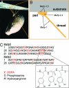
Fig. 1.
Fig. 1.
Biodistribution and amino acid composition of mussel adhesive proteins of M. edulis. (A) Photograph of a mussel attached to a glass surface, showing the byssal threads and adhesive pads (33). (B) The biodistribution of Mefps. Mefp-3 and Mefp-5 are found at the pad–substrate interface. (C) The amino acid sequences of Mefp-3 and Mefp-5, which have the highest known dopa contents, at 21 and 27 mol %, respectively (7, 8). Also shown is the chemical structure of dopa as it appears in the tri-dopa sequence in residues 43–45 of Mefp-5.
Fig. 2.
Fig. 2.
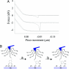
dopa adheres strongly and reversibly to Ti surfaces. (A) Schematic of dopa-functionalized AFM tip and typical single-molecule F–D curves of dopa interacting with a Ti surface. The red and blue traces indicate approach and retraction signals, respectively, in which dopa–Ti adhesive (interaction) force was observed during retraction. The four different force curves were produced from the same dopa-functionalized tip and are displaced vertically for clarity. (B) Histogram (n = 147) of pull-off force values for dopa–Ti obtained with a single AFM tip at a loading rate of 60.0 nN/s. The mean force value was calculated to be 805 ± 131 pN. (C) A linear-log plot of force vs. loading rate for dopa–Ti. Mean forces and standard deviations obtained from a minimum of 30 adhesion events were 847 ± 157 pN (250.0 ± 62.0 nN/s), 805 ± 131 pN (60.0 ± 9.0 nN/s), 744 ± 207 pN (3.50 ± 1.5 nN/s), and 636 ± 151 (0.41 ± 0.15 nN/s).
Fig. 3.
Fig. 3.
Oxidation of dopa reduces adhesion to Ti surfaces. (A) Bimodal force signals from dopa contacting Ti under alkaline conditions (pH 9.7). Selected AFM F–D scans obtained during a 1-h experiment under alkaline conditions (20 mM Tris·HCl, pH 9.7) are shown, with time increasing from the top of the graph to the bottom (top, t = 0; bottom, t = 1 h; 1,800 repetitions total). Detectable force signals were found in ≈10% of the F–D curves obtained. F–D curves exhibiting strong dopa–Ti interactions are shown in red, whereas F–D curves shown in blue exhibited much weaker interactions. The weak force signals appeared only under alkaline conditions and were not observed at neutral pH. (B) Structure-based assignments of the adhesive molecules dopa and dopa–quinone by analysis of 1,800 F–D curves each from two pH conditions (pH 9.7 and 8.3). Bimodal force distributions were measured with averages in a low-force regime [180 ± 60 pN (145 events, pH 9.7) and 206 ± 66 pN (76 events, pH 8.3)] or in a high-force regime [740 ± 110 pN (51 events, pH 9.7) and 759 ± 88 pN (126 events, pH 8.3)]. Considering the pKa (9.2) of the dopa hydroxyl group, the dopa structure is favored over dopa–quinone at low pH, resulting in more frequent detection of high bond force and vice versa at high pH. Considering also the results shown in Fig. 2, which were obtained under a neutral pH condition, where no low-force regime was detected, the strong adhesive interactions are assigned to unoxidized dopa–Ti interaction, whereas the weak adhesive interactions are assigned to the dopa–quinone–Ti interaction. (C) Time trajectory display of the force signals of dopa (red) and dopa–quinone (blue) at pH 9.7 (the time trajectory display at pH 8.3 is available in Fig. 8, which is published as supporting information on the PNAS web site).
Fig. 4.
Fig. 4.
Oxidation of dopa increases adhesion to organic surfaces. (A) Selected F–D curves for interaction of a dopa-modified AFM tip with an organic surface. First, the presence of dopa was confirmed by obtaining a F–D curve at neutral pH on Ti (top curve), showing the expected pull-off force of ≈800 pN. Next, the same tip was allowed to interact with an amine presenting organic surface (Supporting Text) at pH = 9.7, upon which a pull-off force of 2.2 nN was observed (middle curve). The magnitude of the pull-off force is consistent with covalent bond rupture, and subsequent F–D curves (n = 800) failed to show a detectable interaction force (bottom curve). (B) Schematic illustration of covalent bond formation between dopa and amines at the organic surface. The high-magnitude pull-off force, along with lack of subsequent observations of tip–molecule–surface interaction events, suggests that dopa–quinone formed a covalent bond to surface-bound amine, possibly via a Michael addition-type of reaction (–13). The location of the ruptured covalent bond is not known; although, under the conditions of this experiment, it is not expected to re-form, explaining the absence of an adhesion event in subsequent F–D curves.

References

    1. Autumn K., Liang Y. A., Hsieh S. T., Zesch W., Chan W. P., Kenny T. W., Fearing R., Full R. J. Nature. 2000;405:681–685. - PubMed
    1. Chisholm J. R. M., Kelley R. Nature. 2001;409:152. - PubMed
    1. Waite J. H. Integr. Comp. Biol. 2002;42:1172–1180. - PubMed
    1. Waite J. H., Tanzer M. L. Science. 1981;212:1038–1040. - PubMed
    1. Crisp D. J., Walker G., Young G. A., Yule A. B. J. Colloid Interface Sci. 1985;104:40–50.

Publication types