Stability and structure of oligomers of the Alzheimer peptide Abeta16-22: from the dimer to the 32-mer
- PMID: 16920832
- PMCID: PMC1614475
- DOI: 10.1529/biophysj.106.088542
Stability and structure of oligomers of the Alzheimer peptide Abeta16-22: from the dimer to the 32-mer
Abstract
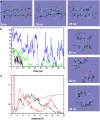
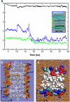
Several neurodegenerative diseases such as Alzheimer's, Parkinson's, and Huntington's diseases are associated with amyloid fibrils formed by different polypeptides. We probe the structure and stability of oligomers of different sizes of the fragment Abeta(16-22) of the Alzheimer beta-amyloid peptide using atomic-detail molecular dynamics simulations with explicit solvent. We find that only large oligomers form a stable beta-sheet aggregate, the minimum nucleus size being of the order of 8-16 peptides. This effect is attributed to better hydrophobic contacts and a better shielding of backbone-backbone hydrogen bonds from the solvent in bigger assemblies. Moreover, the observed stability of beta-sheet aggregates with a different number of layers can be explained on the basis of their solvent-accessible surface area. Depending on the stacking interface between the sheets, we observe straight or twisted structures, which could be linked to the experimentally observed polymorphism of amyloid fibrils. To compare our 32-mer structure to experimental data, we calculate its x-ray diffraction pattern. Good agreement is found between experimentally and theoretically determined reflections, suggesting that our model indeed closely resembles the structures found in vitro.
Figures


Similar articles
-
Oligomerization of amyloid Abeta16-22 peptides using hydrogen bonds and hydrophobicity forces.Biophys J. 2004 Dec;87(6):3657-64. doi: 10.1529/biophysj.104.046839. Epub 2004 Sep 17. Biophys J. 2004. PMID: 15377534 Free PMC article.
-
Molecular dynamics simulations to investigate the structural stability and aggregation behavior of the GGVVIA oligomers derived from amyloid beta peptide.J Biomol Struct Dyn. 2009 Jun;26(6):731-40. doi: 10.1080/07391102.2009.10507285. J Biomol Struct Dyn. 2009. PMID: 19385701
-
Elucidating the Structures of Amyloid Oligomers with Macrocyclic β-Hairpin Peptides: Insights into Alzheimer's Disease and Other Amyloid Diseases.Acc Chem Res. 2018 Mar 20;51(3):706-718. doi: 10.1021/acs.accounts.7b00554. Epub 2018 Mar 6. Acc Chem Res. 2018. PMID: 29508987 Free PMC article. Review.
-
Molecular dynamics simulation of amyloid beta dimer formation.Biophys J. 2004 Oct;87(4):2310-21. doi: 10.1529/biophysj.104.040980. Biophys J. 2004. PMID: 15454432 Free PMC article.
-
Understanding amyloid fibril nucleation and aβ oligomer/drug interactions from computer simulations.Acc Chem Res. 2014 Feb 18;47(2):603-11. doi: 10.1021/ar4002075. Epub 2013 Dec 24. Acc Chem Res. 2014. PMID: 24368046 Review.
Cited by
-
Structure and dynamics of parallel beta-sheets, hydrophobic core, and loops in Alzheimer's A beta fibrils.Biophys J. 2007 May 1;92(9):3032-9. doi: 10.1529/biophysj.106.100404. Epub 2007 Feb 9. Biophys J. 2007. PMID: 17293399 Free PMC article.
-
An Atomistic View of Amyloidogenic Self-assembly: Structure and Dynamics of Heterogeneous Conformational States in the Pre-nucleation Phase.Sci Rep. 2016 Sep 12;6:33156. doi: 10.1038/srep33156. Sci Rep. 2016. PMID: 27616019 Free PMC article.
-
Thermodynamic selection of steric zipper patterns in the amyloid cross-beta spine.PLoS Comput Biol. 2009 Sep;5(9):e1000492. doi: 10.1371/journal.pcbi.1000492. Epub 2009 Sep 4. PLoS Comput Biol. 2009. PMID: 19730673 Free PMC article.
-
Formation and growth of oligomers: a Monte Carlo study of an amyloid tau fragment.PLoS Comput Biol. 2008 Dec;4(12):e1000238. doi: 10.1371/journal.pcbi.1000238. Epub 2008 Dec 5. PLoS Comput Biol. 2008. PMID: 19057640 Free PMC article.
-
Spontaneous formation of twisted Aβ(16-22) fibrils in large-scale molecular-dynamics simulations.Biophys J. 2011 Nov 16;101(10):2493-501. doi: 10.1016/j.bpj.2011.08.042. Epub 2011 Nov 15. Biophys J. 2011. PMID: 22098748 Free PMC article.
References
-
- Dobson, C. 2002. Getting out of shape. Nature. 418:729–730. - PubMed
-
- Dobson, C. 2003. Protein folding and misfolding. Nature. 426:884–890. - PubMed
-
- Selkoe, D. J. 2003. Folding proteins in fatal ways. Nature. 426:900–904. - PubMed
-
- Klein, W. L. 2002. Aβ toxicity in Alzheimer's disease: globular oligomers (ADDLs) as new vaccine and drug targets. Neurochem. Int. 41:345–352. - PubMed
MeSH terms
Substances
LinkOut - more resources
Full Text Sources
Miscellaneous

