. 2006 Sep;142(1):318-32.
doi: 10.1104/pp.106.085415. Epub 2006 Aug 18.

SUMO-conjugating and SUMO-deconjugating enzymes from Arabidopsis

Affiliations

SUMO-conjugating and SUMO-deconjugating enzymes from Arabidopsis

Thomas Colby et al. Plant Physiol. 2006 Sep.

Abstract

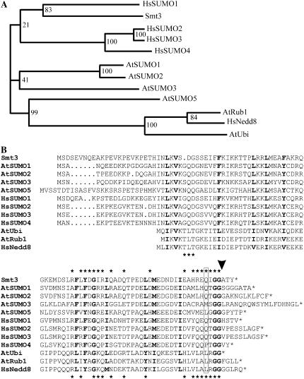
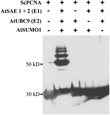
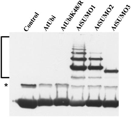
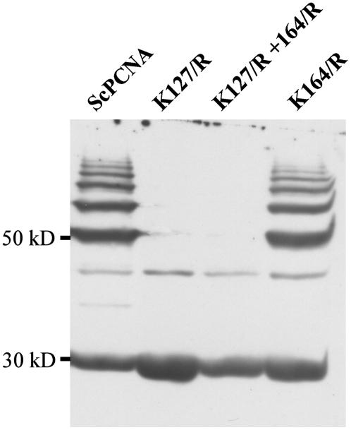
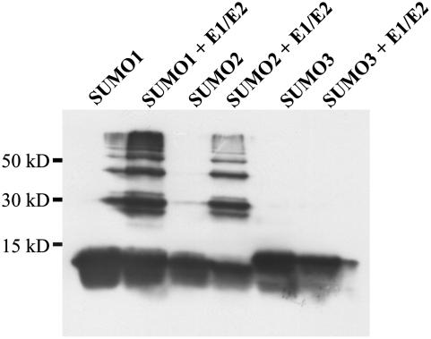
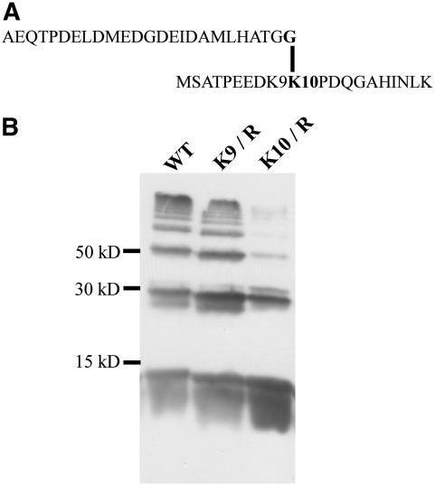
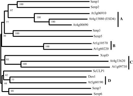
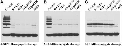
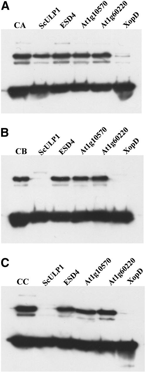
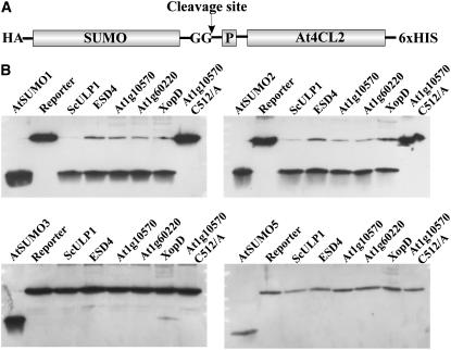
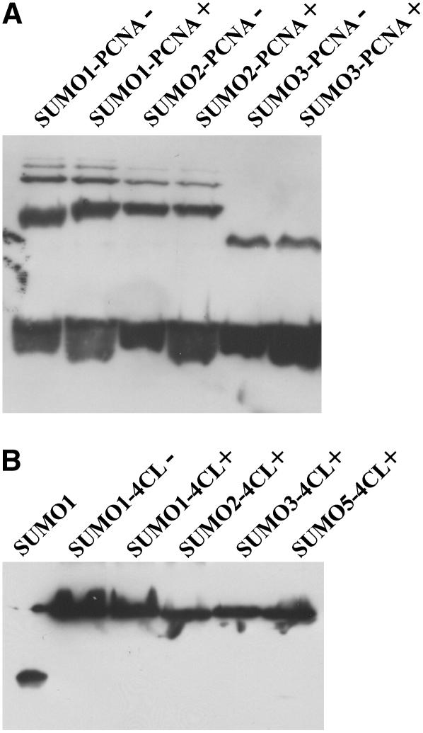
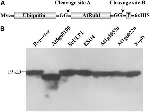
Posttranslational protein modification by the small ubiquitin-like modifier (SUMO) is a highly dynamic and reversible process. To analyze the substrate specificity of SUMO-conjugating and -deconjugating enzymes from Arabidopsis (Arabidopsis thaliana), we reconstituted its SUMOylation cascade in vitro and tested the capacity of this system to conjugate the Arabidopsis SUMO isoforms AtSUMO1, 2, and 3 to the model substrate ScPCNA from yeast (Saccharomyces cerevisiae). This protein contains two in vivo SUMOylated lysine residues, namely K127 and K164. Under in vitro conditions, the Arabidopsis SUMOylation system specifically conjugates all tested SUMO isoforms to lysine-127, but not to lysine-164, of ScPCNA. The SUMO isoforms AtSUMO1 and AtSUMO2, but not AtSUMO3, were found to form polymeric chains on ScPCNA due to a self-SUMOylation process. In a complementary approach, we analyzed both the SUMO isopeptidase activity and the pre-SUMO-processing capacity of the putative Arabidopsis SUMO proteases At1g60220, At1g10570, and At5g60190 using the known SUMO isopeptidases ScULP1, XopD, and ESD4 (At4g15880) as reference enzymes. Interestingly, At5g60190 exhibits no SUMO protease activity but processes the pre-form of Arabidopsis Rub1. The other five enzymes represent SUMO isopeptidases that show different substrate preferences. All these enzymes cleave AtSUMO1 and AtSUMO2 conjugates of ScPCNA, whereas only the putative bacterial virulence factor XopD is able to release AtSUMO3. In addition, all five enzymes cleave pre-AtSUMO1 and pre-AtSUMO2 peptides, but none of the proteins efficiently produce mature AtSUMO3 or AtSUMO5 molecules from their precursors.


Figures

Figure 1.
Figure 1.
Phylogenetic analysis and primary sequence alignment of SUMO and Nedd8 precursors from S. cerevisiae, Arabidopsis, and mammals. A, Proteins were grouped into a phylogram using the ClustalW server at the University of Wageningen (www.bioinformatics.nl/tools/clustalw.html) with standard settings. Bootstrap values (1,000 replicates) are shown at the branches. B, Amino acid residues conserved in all aligned sequences are depicted in bold letters. The C-terminal diglycine motif that is exposed during prepeptide processing is highlighted in bold letters, and the predicted cleavage site is marked by an arrowhead. A glutamine residue known to be important for the interaction of HsSUMO1 with the protease Senp2 is boxed. Asterisks depicted above and below the alignment indicate either Smt3 or HsNedd8 residues that are known to be in direct contact with the proteases ScULP1 or Den1, respectively.
Figure 2.
Figure 2.
SUMOylation of ScPCNA catalyzed by the Arabidopsis SUMOylation cascade. ScPCNA labeled with a Myc tag was isolated from E. coli and directly applied to the gel (lane 1), or incubated with AtSUMO1 in the presences of Arabidopsis SUMO E1 and E2 enzymes (lane 2). In lanes 3 to 5, selected components of the Arabidopsis SUMOylation machinery, as listed in the top part of the figure, were excluded from the reaction mixture. Proteins were separated on a 12% SDS-polyacrylamide gel, and ScPCNA mobility shifts were detected by western-blot analysis using a Myc peptide-specific antiserum.
Figure 3.
Figure 3.
Conjugation of different Arabidopsis SUMO isoforms to ScPCNA. ScPCNA labeled with a Myc tag was purified from E. coli and directly applied to the gel (control in lane 1), or incubated with the Arabidopsis E1 and E2 enzymes and either Arabidopsis ubiquitin (lane 2), the ubiquitin variant K48/R (lane 3), AtSUMO1 (lane 4), AtSUMO2 (lane 5), or AtSUMO3 (lane 6). Proteins were separated on a 12% SDS-polyacrylamide gel, and ScPCNA mobility shifts were detected by western-blot analysis using a Myc peptide-specific antiserum. SUMOylated ScPCNA species are marked by a bracket, and a signal of unclear origin is labeled with an asterisk (*).
Figure 4.
Figure 4.
Identification of SUMOylated lysine residues within ScPCNA. ScPCNA (lane 1) and the ScPCNA protein variants K127/R (lane 2), K127/R + K164/R (lane 3), as well as K164/R (lane 4) were incubated with the Arabidopsis SUMOylation machinery and AtSUMO1. ScPCNA mobility shifts were detected by western-blot analysis using a Myc peptide-specific antiserum.
Figure 5.
Figure 5.
Self-SUMOylation of Arabidopsis SUMO isoforms. The Arabidopsis SUMO isoforms AtSUMO1, AtSUMO2, and AtSUMO3, each labeled with an N-terminal HA tag, were heterologously expressed, purified from E. coli, and either directly applied to the gel or preincubated with a reaction mix containing the Arabidopsis SUMO E1 and E2 enzymes. Proteins were separated on a 15% SDS-polyacrylamide gel, and SUMO mobility shifts were detected by western-blot analysis using a HA peptide-specific antiserum.
Figure 6.
Figure 6.
Analysis of AtSUMO2 multimerization by MS analysis and point mutagenesis. A, Structure of the branched AtSUMO2 peptide identified by LC-MS/MS. B, The Arabidopsis SUMO isoform AtSUMO2 labeled with an N-terminal HA tag (lane 1) and the thereof derived mutant variants K9/R (lane 2) and K10/R (lane 3) were incubated with all components of the Arabidopsis SUMOylation cascade and separated on a 15% SDS-polyacrylamide gel. AtSUMO2 mobility shifts were detected by western-blot analysis using a HA peptide-specific antiserum.
Figure 7.
Figure 7.
Phylogram of ScULP1-like proteins. Arabidopsis proteins that exhibit at least 40% conserved residues when compared with the catalytic domain of ScULP1, as well as Senp1 to 3, Senp5 to 7, Den1, XopD, and ScULP1, were grouped into a phylogenetic tree using the ClustalW server at the University of Wageningen (www.bioinformatics.nl/tools/clustalw.html) with standard settings. Bootstrap values (1,000 replicates) are shown at the branches.
Figure 8.
Figure 8.
SUMO isopeptidase activity and substrate specificity of cysteine proteases from Arabidopsis, yeast, and X. campestris. SUMO isopeptidase activity of ScULP1, ESD4, At1g10570, At1g60220, and XopD was determined using in vitro produced AtSUMO1-ScPCNA (A), AtSUMO2-ScPCNA (B), and AtSUMO3-ScPCNA (C) conjugates. Protease-treated samples were applied as depicted. As a reference, untreated conjugates were always applied to the first lane of the respective gels. Proteins were separated on a 12% SDS-polyacrylamide gel, and cleavage of SUMO-ScPCNA conjugates was detected by western-blot analysis using a Myc peptide-specific antiserum.
Figure 9.
Figure 9.
Substrate quality-determining amino acid residues of AtSUMO3-ScPCNA conjugates. ScPCNA was conjugated in vitro with either AtSUMO3 wild-type protein (A; lane CA), the AtSUMO3 variant M90/Q (B; lane CB), or the AtSUMO3 triple mutant A89/H + M90/Q + S91/T (C; lane CC). Conjugates were treated with either ScULP1 (lanes 2), ESD4 (lanes 3), At1g10570 (lanes 4), At1g60220 (lanes 5), or XopD (lanes 6). Proteins were separated on a 12% SDS-polyacrylamide gel, and cleavage of AtSUMO3-ScPCNA conjugates was detected by western-blot analysis using a Myc peptide-specific antiserum.
Figure 10.
Figure 10.
Pre-SUMO-processing activity and substrate specificity of cysteine proteases from Arabidopsis, yeast, and X. campestris. A, Structure of the reporter construct that was used to detect pre-SUMO-processing activity. The diglycine motif exposed in the mature SUMO molecule is depicted, and the small SUMO prepeptide is labeled with P. The expected site of protease cleavage is marked by an arrow. B, Pre-SUMO-processing activity of ScULP1 (lanes 3), ESD4 (lanes 4), At1g10570 (lanes 5), At1g60220 (lanes 6), XopD (lanes 7), and At1g10570 Cys-512/Ala (lanes 8) was determined using preAtSUMO1-4CL2, preAtSUMO2-4CL, preAtSUMO3-4CL, and preAtSUMO5-4CL reporter constructs. Mature SUMO isoforms isolated from E. coli (lanes 1) and untreated reporter fusions (lanes 2) were loaded in each case as reference probes. Proteins were separated on a 15% SDS-polyacrylamide gel, and cleavage of pre-SUMO reporter constructs was detected by western-blot analysis using a HA peptide-specific antiserum.
Figure 11.
Figure 11.
Analysis of SUMO isopeptidase and pre-SUMO-processing activity of At5g60190. A, The SUMO isopeptidase model substrates AtSUMO1-ScPCNA, AtSUMO2-ScPCNA, and AtSUMO3-ScPCNA were analyzed before (−) and after incubation (+) with At5g60190. Proteins were separated on a 12% SDS-polyacrylamide gel, and SUMO-ScPCNA conjugates were detected by western-blot analysis using a Myc peptide-specific antiserum. B, The pre-SUMO-4CL reporter constructs were incubated with At5g60190 as depicted. As reference probes, AtSUMO1 (lane 1) and the untreated AtSUMO1-4CL protein (lane 2) were applied. Proteins were separated on a 15% SDS-polyacrylamide gel, and the pre-SUMO reporter constructs were detected by western-blot analysis using a HA peptide-specific antiserum.
Figure 12.
Figure 12.
Pre-Rub1 cleavage activity and substrate specificity of At1g60190. A, Structure of the reporter construct that was used to detect pre-AtRub1-processing activity. The reporter is based on a naturally occurring ubiquitin-pre-AtRub1 protein (At1g31340) that was fused with an N-terminal Myc tag and a C-terminal hexa-His tag. Two possible sites of protease cleavage are marked by arrows. The diglycine motifs that are expected to be exposed in the mature molecules are depicted, and the small prepeptide following AtRub1 is labeled with P. B, Pre-AtRub1-processing activity of At5g60190 (lane 2), ScULP1 (lane 3), ESD4 (lane 4), At1g10570 (lane 5), At1g60220 (lane 6), and XopD (lane 7) was determined. Proteins were separated on a 15% SDS-polyacrylamide gel, and cleavage of the reporter construct was detected by western-blot analysis using a Myc peptide-specific antiserum.

Similar articles

Cited by

References

    1. Altschul SF, Gish W, Miller W, Myers EW, Lipman DJ (1990) Basic local alignment search tool. J Mol Biol 215: 403–410 - PubMed
    1. Bayer P, Arndt A, Metzger S, Mahajan R, Melchior F, Jaenicke R, Becker J (1998) Structure determination of the small ubiquitin-related modifier SUMO-1. J Mol Biol 280: 275–286 - PubMed
    1. Bernier-Villamor V, Sampson DA, Matunis MJ, Lima CD (2002) Structural basis for E2-mediated SUMO conjugation revealed by a complex between ubiquitin-conjugating enzyme Ubc9 and RanGAP1. Cell 108: 345–356 - PubMed
    1. Bohren KM, Nadkarni V, Song JH, Gabbay KH, Owerbach D (2004) A M55V polymorphism in a novel SUMO gene (SUMO-4) differentially activates heat shock transcription factors and is associated with susceptibility to type I diabetes mellitus. J Biol Chem 279: 27233–27238 - PubMed
    1. Bradford MM (1976) A rapid and sensitive method for the quantitation of microgram quantities of protein utilizing the principle of protein-dye binding. Anal Biochem 72: 248–254 - PubMed

Publication types

MeSH terms

Substances

Associated data