. 2006 Sep 1;177(5):2880-7.
doi: 10.4049/jimmunol.177.5.2880.

Primitive lymphoid progenitors in bone marrow with T lineage reconstituting potential

Affiliations

Primitive lymphoid progenitors in bone marrow with T lineage reconstituting potential

S Scott Perry et al. J Immunol. .

Abstract

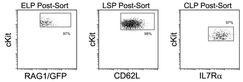
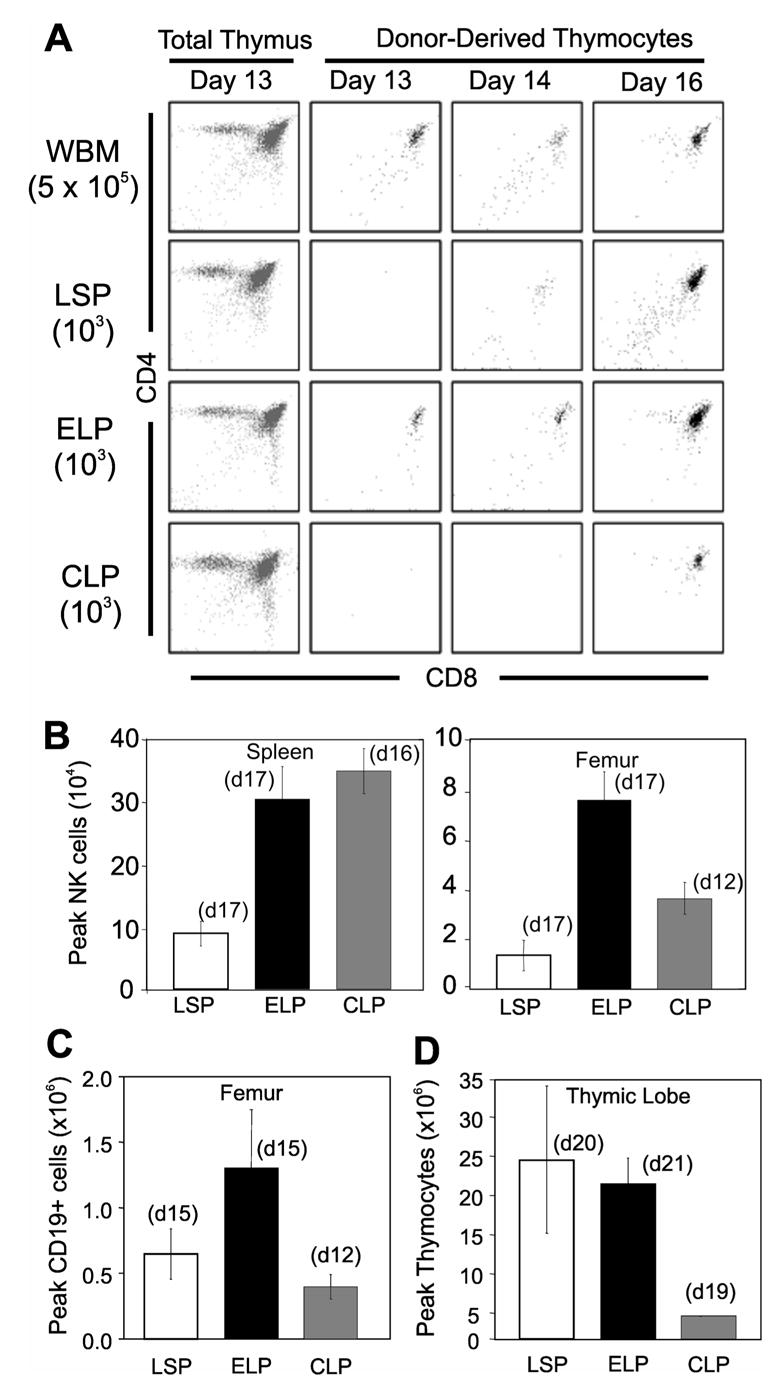
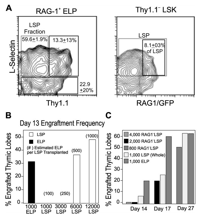
Multiple subsets of the bone marrow contain T cell precursors, but it remains unclear which is most likely to replenish the adult thymus. Therefore, RAG-1+ early lymphoid progenitors (RAG-1+ ELP), and CD62L/L-selectin+ progenitors (LSP), as well as common lymphoid progenitors from C57BL6-Thy1.1-RAG-1/GFP mouse bone marrow were directly compared in transplantation assays. The two c-Kit(high) populations vigorously regenerated the thymus and were superior to common lymphoid progenitors in magnitude and frequency of thymic reconstitution. Regeneration was much faster than the 22 days described for transplanted stem cells, and RAG-1+ ELP produced small numbers of lymphocytes within 13 days. As previously reported, LSP were biased to a T cell fate, but this was not the case for RAG-1+ ELP. Although RAG-1+ ELP and LSP had reduced myeloid potential, they were both effective progenitors for T lymphocytes and NK cells. The LSP subset overlapped with and included most RAG-1+ ELP and many RAG-1- TdT+ ELP. LSP and RAG-1+ ELP were both present in the peripheral circulation, but RAG-1+ ELP had no exact counterpart among immature thymocytes. The most primitive of thymocytes were similar to Lin- c-Kit(high) L-selectin+ TdT+ RAG-1- progenitors present in the marrow, suggesting that this population is normally important for sustaining the adult thymus.


Figures

Fig. 1
Fig. 1. Isolation of three categories of lymphoid progenitors
As detailed in the Materials & Methods, bone marrow suspensions were depleted of lineage positive cells and then sorted as early lymphoid progenitors (ELP; Lin Sca1+ cKitHi RAG-1/GFP+), L-selectin+ progenitors (LSP; Lin Sca1+ cKitHi Thy1.1 L-selectin+) and common lymphoid progenitors (CLP; Lin Sca1Lo cKitLo Thy1.1 IL7R+). All parameters matched the originally described characteristics of these progenitors, and typical post-sort purities are shown. In some experiments, cells were subjected to two rounds of sorting and biological activities were indistinguishable.
Fig. 2
Fig. 2. Lineage potentials of defined progenitor populations
(A) Representative plots of thymic engraftment from i.v. transplants of either 5x10⁵ whole bone marrow leukocytes (WBM) or 103 sorted progenitors. The far left panels show how total thymocytes recover by 13 days post transplant. The remaining dot plots are gated on donor type cells and show how transplanted progenitors generate thymocytes over time. Similar results were seen in experiments that utilized >80 thymic lobes for each transplanted population.Peak contributions of donor cells to NK (B), B cell (C), and thymocyte (D) lineages were evaluated in the indicated tissues. Days at which peak engraftment was achieved per population transplanted are noted in parentheses above each bar. Averages were taken from 5 sorting experiments for LSP, 5 for ELP, and 3 for CLP. Error bars indicate SEM.
Fig. 3
Fig. 3. Kinetics and frequencies of thymocyte engraftment
(A- left panel) Kinetic summary of early thymocytes developing per engrafted lobe from transplants of sorted LSP (squares), ELP (diamonds) or CLP (circles) as described for panel A. Each symbol represents average engraftment from 16–64 lobes analyzed at that time point. (Right panel) The X axis in panel B is expanded to show details of thymic engraftment at early time points. Short-term engraftment from transplantation of 5x10⁵ WBM is given for comparison (triangles). (B) Percentages of lobes containing at least 105 donor thymocytes in the same experiments are shown. (C) Limiting dilution analysis of thymic progenitor frequencies in the LSP and ELP populations. The symbols represent percentages of thymic lobes (minimum n = 8 per dose) containing greater than 105 donor-derived thymocytes on day 17. Lines are best fit of data combined from two independent sorting experiments. Active progenitor frequencies as estimated by limiting dilution analysis for each population are indicated on the x-axis.
Fig. 4
Fig. 4. Overlap between ELP and LSP populations
Six color flow cytometric analysis was used to determine the degree of coincidence between ELP and LSP marrow fractions. (A) The left panel shows that 60% of RAG-1+ ELP express L-selectin and lack Thy 1.1, while an additional 13% of these progenitors display low levels of Thy1.1 (right box). The right panel demonstrates that 8% of LSP (large box) express RAG-1/GFP (small box). The percentages represent means ±SEM from 6 independent experiments. (B) Increasing numbers of progenitors were transplanted to assess competency for rapid regeneration. Thymic lobes containing at least 105 donor thymocytes 13 days post i.v. transplant of ELP (filled bars) and LSP (open bars) are shown. Numbers in parentheses are estimates of the ELP included at each dose of LSP administered, as predicted from the overlap between these populations described in Figure 4 below. A minimum of 10 thymic lobes were analyzed at each transplant dose. (C) T progenitor activity associated with RAG-1 LSP. Percentages are given for thymic lobes with at least 105 donor thymocytes after transplant with LSP depleted of RAG-1+ cells (RAG-1 LSP). Transplantation results with unseparated LSP and ELP (open and gray bars, respectively) are given for reference. Data in C represents two independent experiments, the smaller number of thymic lobes studied could explain the different in frequency of ELP thymic engraftment seen from B and C.
Fig. 5
Fig. 5. Characteristics of progenitors in blood and thymus
(A) A flow cytometry analysis is shown for Lin Thy1.1 cells present in peripheral blood. The left panel resolves LSK (R1) and c-kitLo Sca-1+ (R2) subsets. The R1 population is further separated according to L-selectin and RAG-1/GFP in the middle panel to show RAG-1+ ELP and L-selectin+ LSP. In addition, c-KitLo IL-7Rα + CLP are gated from the R2 subset and illustrated in the far right panel. (B) DN1 thymocytes prepared as described in Materials & Methods were analyzed with respect to c-kit density and intracellular TdT. (C) In two separate experiments, purified bone marrow progenitors were stained for TdT. Average frequencies TdT+ cells relative to negative controls are shown. (D) The thymus DN subsets are resolved in the dot plot and used to set gates for analysis of RAG-1/GFP expression (histograms). (E) RAG-1/GFP is not expressed by DN1 thymocytes with high levels of c-Kit (left panel). The two gated DN1 subsets were placed in fetal thymic organ cultures and only the c-KitHi subsets (R1) generated double positive T lineage lymphocytes (middle and right panels).
Fig. 6
Fig. 6. Relationship between progenitors within the LSK fraction of bone marrow
Gating and resolution of progenitor populations was performed as illustrated in Figure 4A. The relative incidences among total marrow nucleated cells are depicted with circles of proportional size. The degree that subsets have overlapping properties is also indicated.

References

    1. Scollay R, Smith J, Stauffer V. Dynamics of early T cells: prothymocyte migration and proliferation in the adult mouse thymus. Immunol Rev. 1986;91:129–157. - PubMed
    1. Foss DL, Donskoy E, Goldschneider I. The importation of hematogenous precursors by the thymus is a gated phenomenon in normal adult mice. J Exp Med. 2001;193:365–374. - PMC - PubMed
    1. Bhandoola A, Sambandam A. From stem cell to T cell: one route or many? Nat Rev Immunol. 2006;6:117–126. - PubMed
    1. Huang J, Garrett KP, Pelayo R, Zúñiga-Pflücker JC, Petrie HT, Kincade PW. Propensity of adult lymphoid progenitors to progress to DN2/3 stage thymocytes with Notch receptor ligation. J Immunol. 2005;175:4858–4865. - PMC - PubMed
    1. Balciunaite G, Ceredig R, Fehling HJ, Zúñiga-Pflücker JC, Rolink AG. The role of Notch and IL-7 signaling in early thymocyte proliferation and differentiation. Eur J Immunol. 2005;35:1292–1300. - PubMed

Publication types