. 2006 Sep 1;20(17):2410-20.
doi: 10.1101/gad.1454006. Epub 2006 Aug 18.

Emi1 stably binds and inhibits the anaphase-promoting complex/cyclosome as a pseudosubstrate inhibitor

Affiliations

Emi1 stably binds and inhibits the anaphase-promoting complex/cyclosome as a pseudosubstrate inhibitor

Julie J Miller et al. Genes Dev. .

Abstract

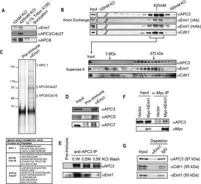
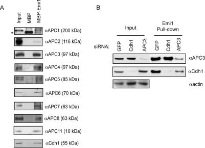
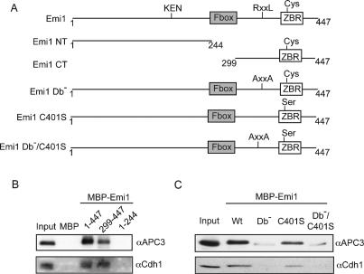
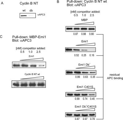
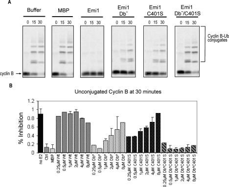
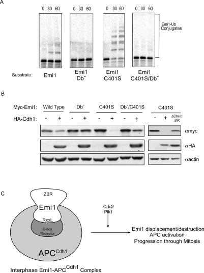
The periodic destruction of mitotic cyclins is triggered by the activation of the anaphase-promoting complex/cyclosome (APC/C) in mitosis. Although the ability of the APC/C to recognize destruction box (D-box) substrates oscillates throughout the cell cycle, the mechanism regulating APC/C binding to D-box substrates remains unclear. Here, we show that the APC/C inhibitor Emi1 tightly binds both the APC/C and its Cdh1 activator, binds to the D-box receptor site on the APC/C(Cdh1), and competes with APC/C substrates for D-box binding. Emi1 itself contains a conserved C-terminal D-box, which provides APC/C-binding affinity, and a conserved zinc-binding region (ZBR), which antagonizes APC/C E3 ligase activity independent of tight APC binding. Mutation of the ZBR converts Emi1 into a D-box-dependent APC/C substrate. The identification of a direct Emi1-APC/C complex further explains how Emi1 functions as a stabilizing factor for cyclin accumulation and the need to destroy Emi1 for APC/C activation in mitosis. The combination of a degron/E3 recognition site and an anti-ligase function in Emi1 suggests a general model for how E3 substrates evolve to become pseudosubstrate inhibitors.


Figures

Figure 1.
Figure 1.
Emi1 associates with the nuclear form of the APC. (A) Emi1 and the APC/C are coextracted from the high-salt fraction of HeLa cells. HeLa cells were extracted by sequential treatments of nitrogen decompression, 420 mM KCl, 0.1% Triton X-100, and sonication to produce fractions as described in Materials and Methods. Fractions were examined by immunoblot analysis for Emi1 and APC/C subunits (APC3/Cdc27, APC8). (B) Emi1, Cdh1, and APC/C cofractionate during ion exchange and gel filtration chromatography. The nuclear fraction described in A was further fractionated by anion exchange (upper panel) chromatography and subsequently separated by gel filtration (lower panel). (C) Emi1 and APC/C coprecipitate from purified fractions. The fractions indicated in B were pooled and immunoprecipitated with anti-Emi1 antibodies and resolved by SDS-PAGE and silver stained. Excised bands were identified by mass spectrometry. APC/C subunits identified by mass spectromety are indicated by arrows. (D) Anti-Emi1 antibody immunoprecipitates were also examined by immunoblot analysis for Emi1 and APC/C subunits, APC3 (Cdc27), APC5, and APC7. (E) Anti-APC3/Cdc27 efficiently coprecipitates Emi1 from nuclear fractions. Immunoprecipitates were washed with 0.1, 0.25, or 0.5 M KCl; separated by SDS-PAGE; and examined by immunoblot analysis for Cdc27 and Emi1. (F) APC/C subunits associate with expressed epitope-tagged Emi1. HeLa cells were transfected with a construct expressing epitope-tagged Myc-hEmi1 or empty vector. Cell extracts were prepared and anti-Myc precipitates were examined by immunoblot analysis for the presence of Emi1 and APC/C subunits. (G) Immunodepletion of asynchronous nuclear extract by anti-Emi1 antibody results in the removal of the majority of APC/C and Cdh1. Nuclear extracts were incubated with anti-Emi1 or control antibody. Equal amounts of input and flow-through (nonbound) samples were examined by immunoblotting.
Figure 2.
Figure 2.
Immobilized Emi1 can bind to the APC/CCdh1 in an “APC/C capture” assay. (A) Recombinant Emi1 protein immobilized on Sepharose beads (see Materials and Methods) was incubated with HeLa cell extracts, and precipitates were analyzed for APC/C binding by immunoblot analysis. The approximate molecular weight at which the indicated subunit migrates is indicated in parentheses. (B) Emi1 binds to the APC/C in the absence of Cdh1. Emi1 beads were incubated with extracts made from HeLa cells transfected with siRNA targeting Cdh1 or APC3 for 48 h.
Figure 3.
Figure 3.
The APC/C binds to the C-terminal APC/C inhibitory domain of Emi1 via its conserved D-box. (A) Emi1 protein schematic, key features, and variant proteins. (B) The Emi1 C-terminal domain binds to the APC/C and Cdh1. Capture of the APC/C using recombinant MBP–Emi1 N terminus (1–244) or C terminus (299–447). (C) Efficient Emi1 binding to the APC/C and Cdh1 requires the C-terminal D-box. Capture of APC/C using MBP–Emi1 D-box mutant (Db), MBP–Emi1 ZBR mutant (C401S), or double mutant (Db/C401S).
Figure 4.
Figure 4.
Emi1 can compete with substrate binding in D-boxand ZBR-dependent manner. (A) The normal N-terminal D-box region of cyclin B1 binds specifically to the APC/C. Recombinant N terminus of cyclin B1 (residues 1–100) or a variant containing a mutated D-box was immobilized on CNBr-activated Sepharose, incubated with cell extracts, and analyzed for APC/C binding with anti-Cdc27 antibody. (B) Emi1 competes with the ability of the cyclin B1 D-box region to bind the APC/ C. Recombinant wild-type or mutant MBP–Emi1 or MBP (0.5, 1, or 2.5 nmol) was added to 1 nmol bead-bound cyclin B1 and incubated with HeLa cell extracts as described in A. The amount of APC/C remaining bound to Cyclin B beads is indicated below, expressed as a percentage of control (no Emi1 addition) sample. Values represent mean of two experiments. (C) Cyclin B1 does not compete efficiently with Emi1 for APC/C binding. N terminus of cyclin B1 or MBP–Emi1 was added at doses of 0.5, 1.0, or 2.5 nmol to capture assay using 1 nmol of bead-bound MBP–Emi1.
Figure 5.
Figure 5.
Emi1’s APC/C inhibitory function requires both the conserved D-box and ZBR functions. (A) APC/ CCdh1-dependent in vitro ubiquitination assays, using radiolabeled, in vitro translated cyclin B1 protein as a substrate, were incubated for the indicated times (0, 15, 30 min), and the formation of ubiquitinated cyclin B was visualized by autoradiography of SDS–polyacryl-amide gels. Wild-type, D-box mutant (Db), ZBR mutant (C401S), and D-box, ZBR double-mutant Emi1 was added to the reaction mixture and assayed for the ability to inhibit cyclin B ubiquitination. (B) Quantitation of in vitro APC/C activity showing the amount of cyclin B remaining unconjugated at 30 min for a range of doses of Emi1 variants.
Figure 6.
Figure 6.
Loss of IBR/ZBR function in Emi1 converts the protein from APC/C inhibitor to APC/C substrate. (A) Standard APC/C-dependent ubiquitination assays, using radiolabled, in vitro translated C-terminal Emi1 wild-type or mutant protein as substrate. Reactions were incubated for 60 min, and the formation of ubiquitinated Emi1 was visualized by autoradiography of SDS–polyacrylamide gels. (Db) D-box mutant. (B) Cdh1 over-expression reduces levels of cotransfected Emi1 C401S/ZBR. HA-Cdh1 and Myc-Emi1 variants were cotransfected into HEK293T cells. Lysates were harvested and Emi1 levels detected by anti-Myc immunoblot. A mutant version of Cdh1 lacking the C-terminal IR dipeptide and the C-box (ΔCboxΔIR) was used as a control. (C) A model for evolving APC/C substrates and inhibitors. Emi1 protein binds to the APC/C in a D-box-dependent manner to inhibit APC/C activity through G2 and early mitosis, permitting cyclin accumulation. The ZBR motif of Emi1 contributes to inhibition by blocking substrate access to the APC/C, as well as providing an additional inhibitory function that prevents the ubiquitination of substrates and Emi1 itself. The combination of D-box and ZBR allows Emi1 to compete with APC/C substrates for APC/C binding. Phosphorylation of Emi1 by Cdc2 and Plk1, potentially in conjunction with other events, results in the displacement and proteolytic destruction of Emi1, allowing for APC/C activation and progression through later stages of mitosis.

References

    1. Bashir T., Dorrello N.V., Amador V., Guardavaccaro D., Pagano M. Control of the SCF(Skp2–Cks1) ubiquitin ligase by the APC/C(Cdh1) ubiquitin ligase. Nature. 2004;428:190–193. - PubMed
    1. Burton J.L., Solomon M.J. Hsl1p, a Swe1p inhibitor, is degraded via the anaphase-promoting complex. Mol. Cell. Biol. 2000;20:4614–4625. - PMC - PubMed
    1. Burton J.L., Tsakraklides V., Solomon M.J. Assembly of an APC–Cdh1–substrate complex is stimulated by engagement of a destruction box. Mol. Cell. 2005;18:533–542. - PubMed
    1. Carroll C.W., Enquist-Newman M., Morgan D.O. The APC subunit Doc1 promotes recognition of the substrate destruction box. Curr. Biol. 2005;15:11–18. - PubMed
    1. Castro A., Bernis C., Vigneron S., Labbe J.C., Lorca T. The anaphase-promoting complex: A key factor in the regulation of cell cycle. Oncogene. 2005;24:314–325. - PubMed

Publication types

MeSH terms