Review
. 2006 Aug;3(6):519-32.
doi: 10.1513/pats.200603-086MS.

State of the Art. A structural and functional assessment of the lung via multidetector-row computed tomography: phenotyping chronic obstructive pulmonary disease

Affiliations
Review

State of the Art. A structural and functional assessment of the lung via multidetector-row computed tomography: phenotyping chronic obstructive pulmonary disease

Eric A Hoffman et al. Proc Am Thorac Soc. 2006 Aug.

Abstract

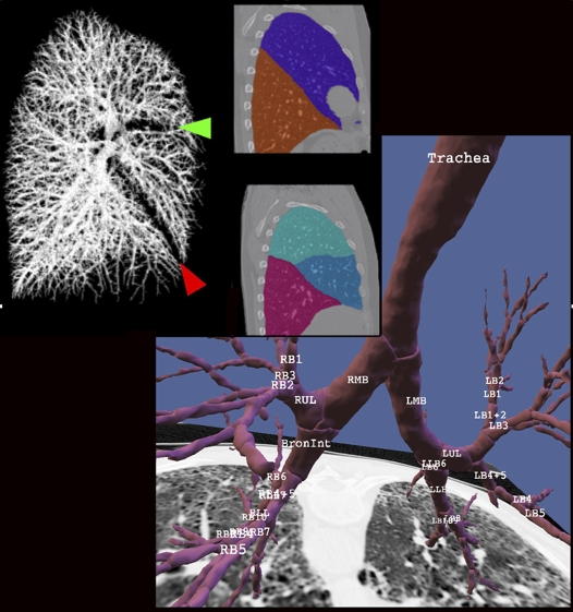
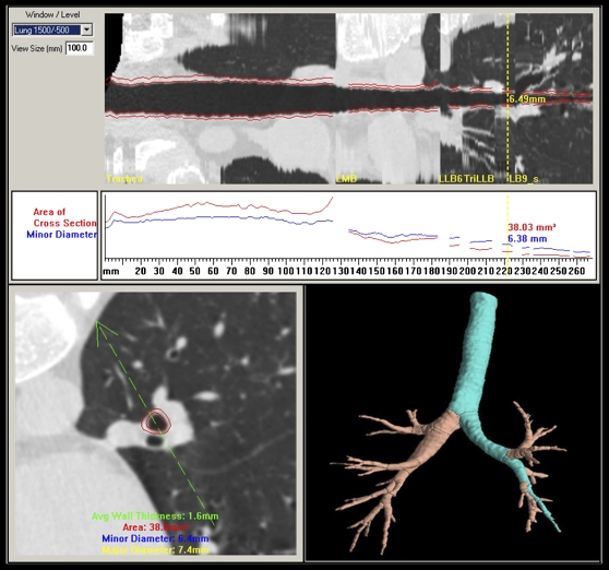
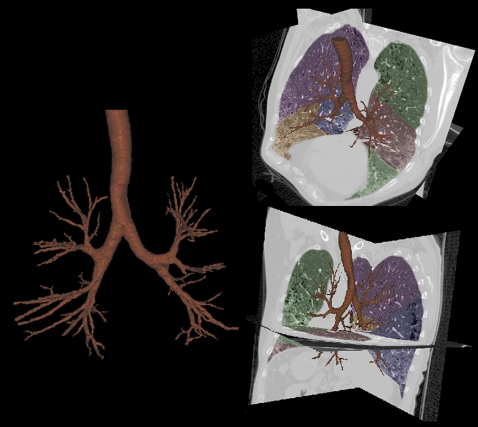
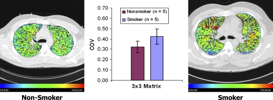
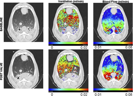
With advances in multidetector-row computed tomography (MDCT), it is now possible to image the lung in 10 s or less and accurately extract the lungs, lobes, and airway tree to the fifth- through seventh-generation bronchi and to regionally characterize lung density, texture, ventilation, and perfusion. These methods are now being used to phenotype the lung in health and disease and to gain insights into the etiology of pathologic processes. This article outlines the application of these methodologies with specific emphasis on chronic obstructive pulmonary disease. We demonstrate the use of our methods for assessing regional ventilation and perfusion and demonstrate early data that show, in a sheep model, a regionally intact hypoxic pulmonary vasoconstrictor (HPV) response with an apparent inhibition of HPV regionally in the presence of inflammation. We present the hypothesis that, in subjects with pulmonary emphysema, one major contributing factor leading to parenchymal destruction is the lack of a regional blunting of HPV when the regional hypoxia is related to regional inflammatory events (bronchiolitis or alveolar flooding). If maintaining adequate blood flow to inflamed lung regions is critical to the nondestructive resolution of inflammatory events, the pathologic condition whereby HPV is sustained in regions of inflammation would likely have its greatest effect in the lung apices where blood flow is already reduced in the upright body posture.


Figures

<b>Figure 1.</b>
Figure 1.
Results of vascular (upper left), lobe (middle), and airway (lower right) segmentation. After the airways are identified (segmented) and the centerline and branchpoints are identified, then the airway tree is automatically labeled. The lobar fissures are identified by the geometry of the segmented blood vessels as shown by the red and green arrowheads shown in the upper left panel. L = left, R = right, B = broncus, UL = upper lobe, M = middle lobe, LL = lower lobe.
<b>Figure 2.</b>
Figure 2.
Once the three-dimensional airway tree has been identified and labeled, paths can be identified and straightened so as to provide luminal and wall dimensions measured as a function of the distance along the path and perpendicular to the local long axis. The airway cross-section corresponding to the yellow vertical line in the upper panel is shown in the lower left panel. The green arrow in the lower left panel can be rotated about the centerline of the airway to alter the cut plane shown in the straightened airway presented in the upper panel.
<b>Figure 3.</b>
Figure 3.
Demonstration of the ability to extract a detailed airway tree from computed tomography (CT) scans of a patient with significant mixed-lung pathology. The images show emphysema, honeycombing, traction bronchiectasis, and fibrosis.
<b>Figure 4.</b>
Figure 4.
A normal nonsmoker and GOLD 0 smoker showing considerable increase in parenchymal perfusion heterogeneity. When sampling at increasingly coarse sample sizes, the difference in the coefficients of variation (COV) between the two groups disappears when the region-of-interest size is larger that 3 × 3 voxels (∼ 1.2 mm on a side, about one-fifth to one-tenth the size of an adult ascinus) (174). Color coding ranges from 0 (blue) to 15 (red) ml/min.
<b>Figure 5.</b>
Figure 5.
Before (baseline; top row) and after (post valve; bottom row) endobronchial valve placement. The left column provides a view of one CT section at the level of the diaphragm dome. Note the significant dependent pneumonia (A). The center column demonstrates ventilation overlayed in color as assessed by xenon-CT. Note that there is little ventilation in either the baseline or the post valve in the region of the dependent pneumonia. By design, there is a large region where ventilation was eliminated by the valve placement (B). The right column shows a color overlay of the perfusion measurements. Note that in the region (C) coinciding with region B from the central column (no ventilation due to valve placement), there is a regional loss of perfusion, indicating an intact hypoxic pulmonary vasoconstrictor (HPV) response. At the same time, in the region of pneumonia (D) where there is little or no ventilation, blood flow is enhanced after valve placement. Presumably, blood flow shunted away from the valve-based HPV is shunted straight toward the region of inflammation, presumably because the inflammation has served to blunt HPV in this region, leaving this region as the path of least resistance for the blood flow diverted from the region effected by HPV. Thus, this image demonstrates that lung has the ability to locally modulate HPV based on local inflammation.

References

    1. Remy-Jardin M, Edme JL, Boulenguez C, Remy J, Mastora I, Sobaszek A. Longitudinal follow-up study of smoker's lung with thin-section CT in correlation with pulmonary function tests. Radiology 2002;222:261–270. - PubMed
    1. Moonen M, Xu J, Johansson A, Thylen A, Bake B. Effects of lung volume reduction surgery on distribution of ventilation and perfusion. Clin Physiol Funct Imaging 2005;25:152–157. - PubMed
    1. Xu JH, Moonen M, Johansson A, Bake B. Distribution of ventilation-to-perfusion ratios analysed in planar scintigrams of emphysematous patients. Clin Physiol 2000;20:89–94. - PubMed
    1. Cederlund K, Hogberg S, Jorfeldt L, Larsen F, Norman M, Rasmussen E, Tylen U. Lung perfusion scintigraphy prior to lung volume reduction surgery. Acta Radiol 2003;44:246–251. - PubMed
    1. Hunsaker AR, Ingenito EP, Reilly JJ, Costello P. Lung volume reduction surgery for emphysema: correlation of CT and V̇/Q̇ imaging with physiologic mechanisms of improvement in lung function. Radiology 2002;222:491–498. - PubMed

Publication types

MeSH terms