. 2006 Aug 15;54(3):147-59.
doi: 10.1002/glia.20369.

Schwann cell-like differentiation by adult oligodendrocyte precursor cells following engraftment into the demyelinated spinal cord is BMP-dependent

Affiliations

Schwann cell-like differentiation by adult oligodendrocyte precursor cells following engraftment into the demyelinated spinal cord is BMP-dependent

Jason F Talbott et al. Glia. .

Abstract

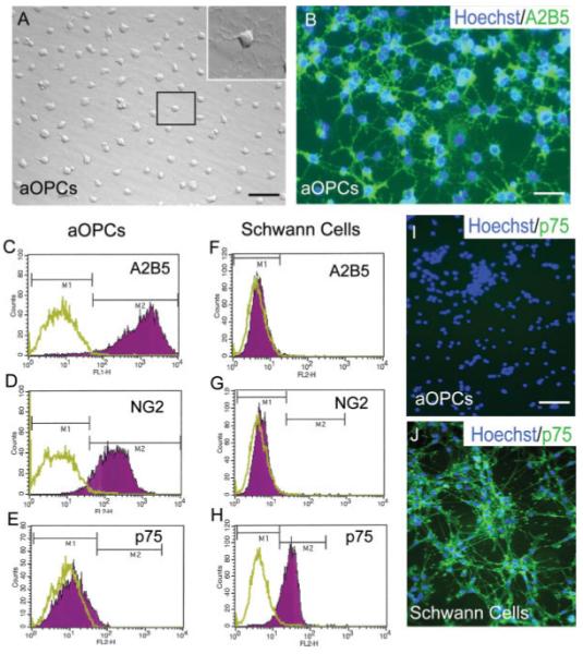
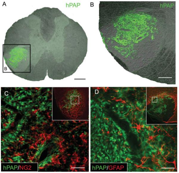
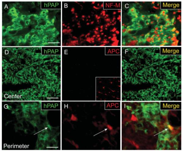
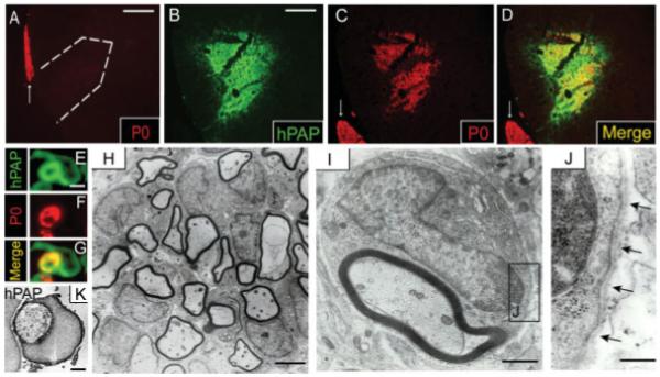
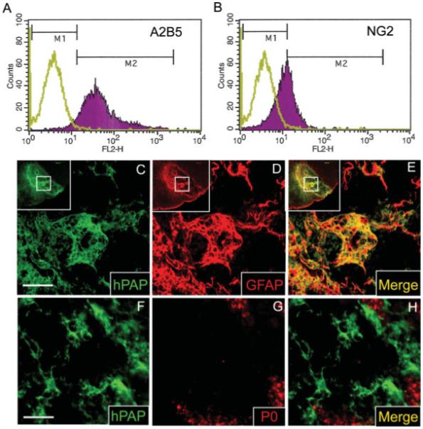
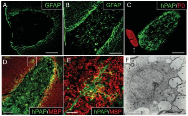
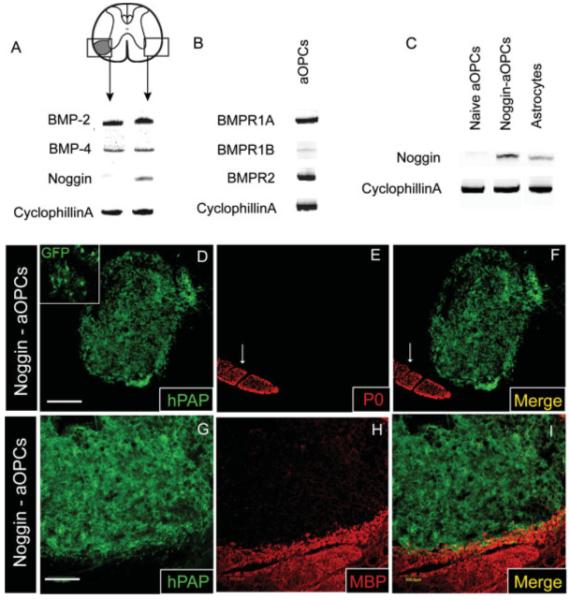
The development of remyelinating strategies designed to enhance recruitment and differentiation of endogenous precursor cells available to a site of demyelination in the adult spinal cord will require a fundamental understanding of the potential for adult spinal cord precursor cells to remyelinate as well as an insight into epigenetic cues that regulate their mobilization and differentiation. The ability of embryonic and postnatal neural precursor cell transplants to remyelinate the adult central nervous system is well documented, while no transplantation studies to date have examined the remyelinating potential of adult spinal-cord-derived oligodendrocyte precursor cells (adult OPCs). In the present study, we demonstrate that, when transplanted subacutely into spinal ethidium bromide/X-irradiated (EB-X) lesions, adult OPCs display a limited capacity for oligodendrocyte remyelination. Interestingly, the glia-free environment of EB lesions promotes engrafted adult OPCs to differentiate primarily into cells with immunophenotypic and ultrastructural characteristics of myelinating Schwann cells (SCs). Astrocytes modulate this potential, as evidenced by the demonstration that SC-like differentiation is blocked when adult OPCs are co-transplanted with astrocytes. We further show that inhibition of bone morphogenetic protein (BMP) signaling through noggin overexpression by engrafted adult OPCs is sufficient to block SC-like differentiation within EB-X lesions. Present data suggest that the macroglial-free environment of acute EB lesions in the ventrolateral funiculus is inhibitory to adult spinal cord-derived OPC differentiation into remyelinating oligodendrocytes, while the presence of BMPs and absence of noggin promotes SC-like differentiation, thereby unmasking a surprising lineage fate for these cells.


Figures

Fig. 1
Fig. 1
Adult oligodendrocyte precursor cell (adult OPC) cultures are not contaminated with SCs. (A) Phase contrast microscopy shows that adult OPCs exhibit several processes emanating from a small round cell body (inset shows higher magnification view of boxed cell). (B) Nearly all cells express A2B5, as demonstrated by immunohistochemistry. FACS analysis shows uniform expression of the glial precursor markers A2B5 (98%) and NG2 (93%) and an absence of p75 expression by adult OPC cultures (CE). In contrast to adult OPCs, SCs do not express A2B5 or NG2 but greater than 95% express p75 (FH). (I) Immunohistochemistry confirms a lack of p75 expression by adult OPCs while SCs demonstrate uniform expression (J). (C–H) The green plot represents unlabeled control cells while the purple plot represents the population of cells labeled with fluorescent antibody. (C–H) Data are representative of three independent experiments. Scale bar = 24 μm (A); 18 μm(B); 28 μm (I, J).
Fig. 2
Fig. 2
Adult OPCs engrafted into ventrolateral funiculus (VLF) ethidium bromide (EB) lesions of the adult rat thoracic spinal cord survive 4 weeks after transplantation and do not differentiate into astrocytes. (A) Nomarsky image of engrafted EB lesions merged with an immunohistochemical stain for the grafted cell marker human placental alkaline phosphatase (hPAP) demonstrates the survival and integration of transplanted cells within VLF EB lesions 4 weeks following transplantation. (B) Higher magnification view of area boxed in (A). hPAP double-labeling reveals that engrafted adult OPCs no longer express the precursor marker NG2 4 weeks after transplantation (C). hPAP double-labeling shows that engrafted adult OPCs do not differentiate significantly into GFAP+ astrocytes 4 weeks after transplantation (D). Insets (C, D) show the entire region of the VLF and the inner box designates the area shown at higher magnification. Scale bar = 250 μm(A); 125 μm (B); 35 μ(C);25 μm(D).
Fig. 3
Fig. 3
Adult OPCs engrafted into EB-X lesions ensheath axons but do not differentiate into mature oligodendrocytes in the center of lesions. hPAP immunohistochemistry for engrafted cells 4 weeks after transplantation demonstrates that transplanted cells exhibit a characteristic ring-like morphology and many of these rings ensheath NF-M+ axons. (AC) Despite their myelin-like formations around axons, double-label immunohistochemistry for hPAP and the mature oligodendrocyte marker APC/CC-1 shows that engrafted cells in the center of EB-X lesions do not differentiate into oligodendrocytes (DF). Inset in (E) shows positive APC/CC-1 staining for oligodendrocytes in the uninjured, contralateral VLF. An occasional hPAP+/APC+ cell (white arrow) was observed in the perimeter of such lesions (GI). Scale bar = 20 μm (A–C);36 μm in (D–F); 16 μm in (G–I).
Fig. 4
Fig. 4
Adult OPCs engrafted into EB-X lesions differentiate into P0-expressing cells ultrastructurally indistinguishable from SCs. The white arrows denote P0+ ventral nerve roots (A,C,D). (A) EB-X lesions do not demonstrate spontaneous SC remyelination 4 weeks postlesion as evidenced by a lack of P0+ cells within lesions. Dashed white line (A) approximates the lesion boundary. Double-label immunohistochemistry shows co-localization between hPAP+ engrafted cells and large patches of P0+ cells throughout VLF EB-X lesions 4 weeks after transplantation (BD). High magnification double-label confocal microscopy of a single hPAP+ cell within an EB-X lesion reveals a Schwann cell-like morphology with P0 expression in the region approximating a myelin ring (EG). (H) Electron microscopy confirmed the presence of numerous Schwann-like cells within adult OPC engrafted EB-X lesions. (I) Higher magnification image of a single Schwann-like cell within an engrafted EB-X lesion revealing ultrastructural similarity to a myelinating SC, including a 1:1 cell-to-axon ratio and a basement membrane surrounding the cell membrane (J). Arrows in (J) denote the basement membrane surrounding the Schwann-like cell. (K) Immuno-electron microscopy shows robust plasmalemmal hPAP expression by an engrafted cell ultrastructurally resembling a SC. Scale bar = 120 μm (A); 80 μm (B–D); 3 μm (E–G); 1 μm (K and I); 3 μm (H); 0.3 μm (J).
Fig. 5
Fig. 5
Glial-restricted precursors (GRPs) immunopurified from the E14 rat spinal cord differentiate into astrocytes rather than SCs 4 weeks following transplantation into EB lesions. Embryonic GRPs express the glial precursor antigens A2B5 and NG2 as assessed by FACS (A, B). In (A, B) the green plot represents unlabeled control cells while the purple plot represents the population of cells labeled with fluorescent antibody. Double-label immunohistochemistry for hPAP and GFAP shows that most GRPs differentiate into astrocytes 4 weeks after engraftment into EB lesions of the ventrolateral spinal cord (CE). (C–E) The insets show the entire region of the VLF and the inner box designates the area shown at higher magnification. Unlike adult OPCs, GRPs do not co-label with the SC myelin protein P0 following engraftment into EB lesions (FH). As evidenced by the presence of some endogenous SC remyelination (G), this particular animal was not irradiated. Similar results were obtained irrespective of whether or not animals received irradiation, with the exception that endogenous SCs were not observed within EB-X lesions (data not shown). Scale bar = 50 μm in (C–E); 28 μm in (F–H).
Fig. 6
Fig. 6
Adult OPCs co-transplanted into EB-X lesions with astrocytes do not differentiate into P0+ SCs. (A) EB is toxic to astrocytes as evidenced by a lack of GFAP+ astrocytes within nontransplanted lesions 4 weeks after EB injection. (B) Many GFAP+ astrocytes are present within astrocyte/adult OPC co-transplanted EB-X lesions. (C) hPAP and P0 double-label immunohistochemistry of astrocyte/adult OPC co-transplanted EB-X lesions reveals a lack of P0+ SC differentiation by hPAP+ adult OPCs. The arrow (C) denotes a P0+ ventral nerve root juxtaposed to the engrafted EB-X lesion. (D) MBP and hPAP co-labeling shows a lack of oligodendrocyte myelin within adult OPC/astrocyte engrafted lesions. Boxed area in (D) is shown at higher magnification in (E). (E) In the perimeter of EB-X lesions occasional adult OPCs appear to differentiate into cells with a mature oligodendrocyte phenotype as evidenced by multiple processes extending out to MBP+ myelin rings. (F) Immunohistochemical evidence of mature oligodendrocyte differentiation by engrafted cells in the perimeter of EB-X lesions is supported by ultrastructural evidence of remyelinating oligodendrocytes in this same area. Thin myelin rings characteristic of remyelinating oligodendrocytes are seen adjacent to a mature oligodendrocyte cell body. Scale bar = 165 μm (A); 83 μm (B); 160 μm (C); 100 μm (D); 20 μm (E); 2 μm (F).
Fig. 7
Fig. 7
Bmp-2 and bmp-4 transcripts are expressed within EB lesions while noggin mRNA is only weakly expressed. Retroviral transduction of adult OPCs with the BMP antagonist noggin is sufficient to block their differentiation into Schwann-like cells following engraftment into EB lesions. White arrows (E, F) denote P0+ ventral nerve roots. (A) RT-PCR analysis of micro-dissected EB lesioned tissue and contralateral uninjured VLF 6 days after EB injection reveals expression of both bmp-2 and bmp-4 mRNA in both demyelinated and uninjured tissue. Noggin mRNA is clearly present in the uninjured VLF tissue but barely detectable in the demyelinated VLF. (B) Adult OPCs are potentially responsive to BMPs as evidenced by their expression of transcripts for bmpr-Ia and bmpr-II. Adult OPCs only weakly express bmpr-Ib. (C) RT-PCR analysis of astrocytes, naïve adult OPCs, and noggin-transduced adult OPCs reveals expression of noggin mRNA in transduced adult OPCs and astrocytes while naïve adult OPCs only weakly express noggin. (DF) Adult OPCs transduced with a retrovirus to express green fluorescent protein (GFP) and noggin fail to differentiate into P0+ Schwann-like cells 4 weeks after engraftment. Upper left inset in (D) shows GFP expression by engrafted cells, confirming the presence of noggin-transduced cells within these EB lesions. (GI) Noggin+ adult OPCs do not differentiate into remyelinating oligodendrocytes as evidenced by a lack of MBP expression within Noggin-adult OPC engrafted EB-X lesions. Scale bar = 250 μm (D–F); 100 μm (G–I).

References

    1. Akiyama Y, Honmou O, Kato T, Uede T, Hashi K, Kocsis JD. Transplantation of clonal neural precursor cells derived from adult human brain establishes functional peripheral myelin in the rat spinal cord. Exp Neurol. 2001;167:27–39. - PubMed
    1. Akiyama Y, Radtke C, Honmou O, Kocsis JD. Remyelination of the spinal cord following intravenous delivery of bone marrow cells. Glia. 2002;39:229–236. - PMC - PubMed
    1. Blakemore WF. Remyelination by Schwann cells of axons demye-linated by intraspinal injection of 6-aminonicotinamide in the rat. J Neurocytol. 1975;4:745–757. - PubMed
    1. Blakemore WF. Limited remyelination of CNS axons by Schwann cells transplanted into the sub-arachnoid space. J Neurol Sci. 1984;64:265–276. - PubMed
    1. Blakemore WF. The case for a central nervous system (CNS) origin for the Schwann cells that remyelinate CNS axons following concurrent loss of oligodendrocytes and astrocytes. Neuropathol Appl Neurobiol. 2005;31:1–10. - PubMed

Publication types

Substances

LinkOut - more resources