. 2006 Aug 29;45(34):10260-9.
doi: 10.1021/bi060490t.

CFTR Expression in human neutrophils and the phagolysosomal chlorination defect in cystic fibrosis

Affiliations

CFTR Expression in human neutrophils and the phagolysosomal chlorination defect in cystic fibrosis

Richard G Painter et al. Biochemistry. .

Abstract

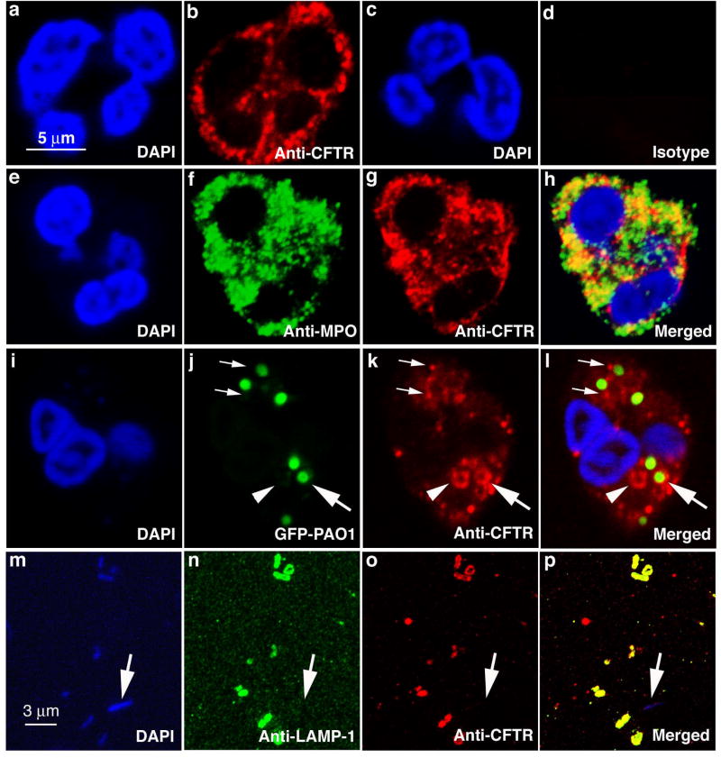
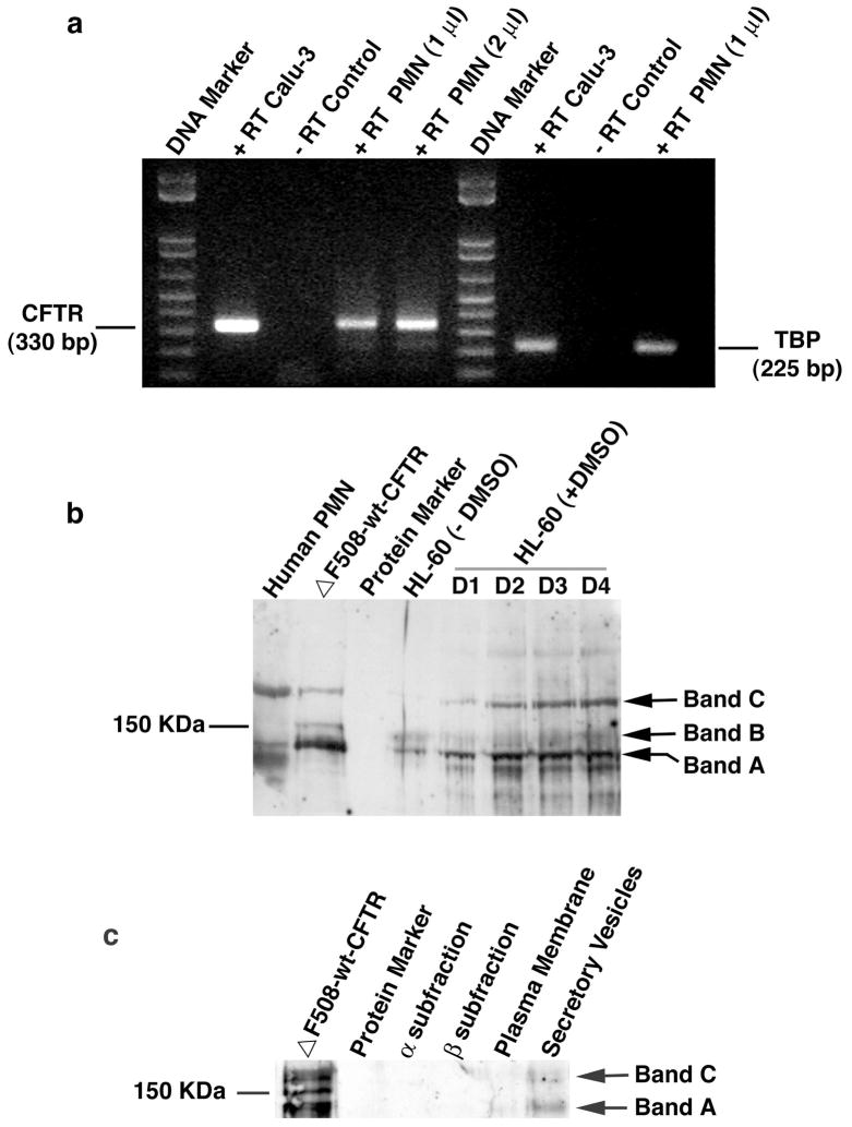
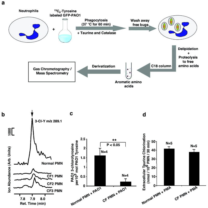
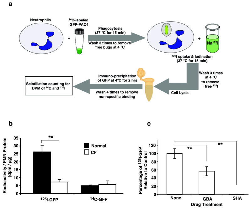
Production of hypochlorous acid (HOCl) in neutrophils, a critical oxidant involved in bacterial killing, requires chloride anions. Because the primary defect of cystic fibrosis (CF) is the loss of chloride transport function of the CF transmembrane conductance regulator (CFTR), we hypothesized that CF neutrophils may be deficient in chlorination of bacterial components due to a limited chloride supply to the phagolysosomal compartment. Multiple approaches, including RT-PCR, immunofluorescence staining, and immunoblotting, were used to demonstrate that CFTR is expressed in resting neutrophils at the mRNA and protein levels. Probing fractions of resting neutrophils isolated by Percoll gradient fractionation and free flow electrophoresis for CFTR revealed its presence exclusively in secretory vesicles. The CFTR chloride channel was also detected in phagolysosomes, a special organelle formed after phagocytosis. Interestingly, HL-60 cells, a human promyelocytic leukemia cell line, upregulated CFTR expresssion when induced to differentiate into neutrophils with DMSO, strongly suggesting its potential role in mature neutrophil function. Analyses by gas chromatography and mass spectrometry (GC-MS) revealed that neutrophils from CF patients had a defect in their ability to chlorinate bacterial proteins from Pseudomonas aeruginosa metabolically prelabeled with [(13)C]-l-tyrosine, unveiling defective intraphagolysosomal HOCl production. In contrast, both normal and CF neutrophils exhibited normal extracellular production of HOCl when stimulated with phorbol ester, indicating that CF neutrophils had the normal ability to produce this oxidant in the extracellular medium. This report provides evidence which suggests that CFTR channel expression in neutrophils and its dysfunction affect neutrophil chlorination of phagocytosed bacteria.


Figures

Figure 1
Figure 1. Localization of CFTR in human neutrophils and phagolysosomes
a & c, DAPI staining of nuclei of human neutrophils. b, anti-CFTR antibody staining of a human neutrophil revealing a punctate staining pattern. d, isotype-matched antibody staining as a control. e–h, double immunofluorescent staining with rabbit anti-human myeloperoxidase antibody (f) and mouse anti-CFTR antibody (g). The merged image (h) to identify co-localization of CFTR and MPO. i–l, CFTR association with phagocytic vacuoles and phagolysosomes bearing ingested green fluorescence protein-expressing Pseudomonas aeruginosa (GFP-PAO1). DAPI staining of a neutrophil with ingested bacteria (i). Phagocytosed GFP-PAO1 (j). Anti-CFTR immunofluorescent staining of a neutrophil with ingested bacteria (k). Association of internalized GFP-PAO1 bacteria with CFTR (l). Large arrows point to a phagolysosome where CFTR is present on the membrane (j–i). Small arrows point to the CFTR-positive staining granules appearing attached to the phagosomes with ingested GFP-PAO1 (j–i). Arrowheads point to a phagocytic vacuole (j–i). m–p, co-localization of CFTR and lysosomal associated membrane protein-1(LAMP-1) in isolated phagolysosomes. DAPI staining of phagolysosomes with ingested non-fluorescent PAO1 which are stained blue (m). LAMP-1 is localized to the phagolysosomes (n). CFTR is present in the phagolysosomal membranes (o). Merged image shows the co-localization of the two proteins (p).
Fig. 2
Fig. 2. CFTR expression in human neutrophils (PMN) and differentiated HL-60 cells induced by DMSO
a) RT-PCR to identify CFTR expression in human neutrophils at the mRNA level. Neutrophils were isolated from whole peripheral blood by Percoll-gradients and further purified by a panning procedure taking advantage of neutrophil adherence to plastic surface. Total RNAs were extracted and cDNAs were obtained by reverse-transcription using the CFTR and TBP specific primers. PCR amplification resulted in a CFTR amplicon of 330 bp and TBP 225 bp. Calu-3 is an epithelial cell line derived from airway submucosal gland, which is known to express high levels of CFTR. − RT or + RT represents PCR in the absence or presence of the corresponding RT product. PMN (1 μl) or PMN (2 μl) indicates PCR with 1 μl or 2 μl of PMN RT product to show the dose-dependence of RT. b) Immunoblotting of CFTR of human neutrophils and differentiated HL-60 cells. Neutrophils or HL-60 cells were denatured by TCA to prevent proteolytic degradation by neutrophil-bearing proteases. 7.5% SDS-PAGE was used to resolve the proteins. After being transferred to a nitrocellulose membrane, the membrane was incubated with the CFTR-specific antibody followed by incubation with the goat anti-mouse IgG conjugated with horseradish peroxidase for chemilluminescent detection. ΔF508-wt-CFTR: wild-type CFTR gene-corrected epithelial cell line derived from a ΔF508 CF patient; HL-60: human promyelocytic leukemia cell line. Bands A, B and C represent the three forms of CFTR (Band A: newly synthesized CFTR; Band B: partially glycosylated CFTR; Band C: fully glycosylated mature CFTR). D1–4 indicates times in days after DMSO treatment. c) Immunoblotting of CFTR in subcellular fractions of normal human neutrophils. MPO-rich α granule fractions, vitamin B-12 binding protein-rich β granules and alkaline phosphatase-rich γ fractions were isolated. The γ fractions were further separated into secretory vesicles (SV) and plasma membrane-derived vesicles (PM) by free flow electrophoresis after treated with neuronamidase. 20–50 × 106 cell equivalents of α and β fractions were TCA-precipitated, and the dissolvable proteins were loaded into each lane. For SV and PM, the dissolvable proteins from ~350 × 106 cell equivalents of TCA-precipitated fractions were loaded, respectively. The CFTR expression in the forms of newly synthesized non-glycosylated protein (Band A) and the mature glycosylated protein (Band C) was predominantly detected in the SV fraction.
Figure 3
Figure 3. Intraphagolysosomal chlorination of bacterial proteins by human neutrophils from normal and CF donors
a, schematic flow diagram of the experimental protocol. Neutrophils (PMN) were incubated with green fluorescence protein-expressing Pseudomonas aeruginosa (GFP-PAO1) which had been metabolically prelabeled with 13C9-L-tyrosine. After 60 min at 37° C, the uningested bacteria were removed by low speed centrifugation and the neutrophils containing ingested PAO1 were analyzed for 13C9-3-chlorotyrosine (13C9-3-Cl-Y) and 13C9-L-tyrosine (13C9-Y) as described in the Materials and Methods using the isotope dilution method. 13C6-3-Cl-Y (20 pmol) and 13C6-Y (50 nmol) were used as internal standards. After derivatization, the samples were analyzed by GC/MS using the selected ion mode. b, Representative GC/MS tracings obtained with normal and CF neutrophils. The 289.1 m/z peak eluting at 7.88 min, obtained by monitoring the ion at m/Z 289.1, is associated with PAO1-derived 3-chlorotyrosine. Normal neutrophils show significantly higher levels of chlorotyrosine relative to that seen from CF neutrophils. CF1, CF2, and CF3 are neutrophils (PMN) from 3 different CF donors. c, Ratios of PAO1-derived 3-chlorotyrosine relative to total PAO1-derived tyrosine levels after one hour of incubation with normal or CF patient neutrophils. Error bars indicate the standard error of the mean. Student’s t-test proved a significant difference between normal and CF PMNs (P<0.05, N=4). d, Chlorination of extracellular taurine by HOCl generated by normal and CF neutrophils induced by phorbol-12-myristate-13-acetate (PMA). Statistically, no significant difference was seen between the two groups.
Figure 4
Figure 4. Intraphagolysosomal iodination of bacterial proteins by human neutrophils from normal and CF donors
a, Schematic depiction of the experimental protocol used to quantitatively measure iodination by neutrophils (PMN) of the green fluorescent protein (GFP) expressed in GFP-expressing Pseudomonas aeruginosa. b, Incorporation of 125I or 14C into GFP immunoprecipitated from neutrophils derived from normal donors (n= 4; closed bars) or donors with CF (n=4; open bars). The recovery of 14C-GFP was similar in both normal and CF cells indicating that the amount of 14C-labeled GFP-PAO1 phagocytosed by neutrophils and its subsequent recovery were statistically identical. In contrast, the 125I content of recovered GFP was about 4.3-fold higher in normal neutrophils than in CF neutrophils. The error bars represent the SEM and the double asterisks represent a P value < 0.05. c, Glybenclamide (GBA), a CFTR channel inhibitor, significantly blocked iodination of bacteria-GFP derived from GFP-PAO1 phagocytosed by normal neutrophils as compared to controls treated with drug vehicle (None) (N=5, P<0.05). In contrast, salicylhydroxamic acid (SHA), an inhibitor of MPO, totally abolished iodination of bacteria-GFP by normal neutrophils relative to controls (N=5, P<0.01). The double asterisks indicate significant differences.

References

    1. Rommens JM, Iannuzzi MC, Kerem B, Drumm ML, Melmer G, Dean M, Rozmahel R, Cole JL, Kennedy D, Hidaka N, et al. Identification of the cystic fibrosis gene: chromosome walking and jumping. Science. 1989;245:1059–1065. - PubMed
    1. Riordan JR, Rommens JM, Kerem B, Alon N, Rozmahel R, Grzelczak Z, Zielenski J, Lok S, Plavsic N, Chou JL, et al. Identification of the cystic fibrosis gene: cloning and characterization of complementary DNA. Science. 1989;245:1066–1073. - PubMed
    1. Welsh MJ, Ramsey BW, Accurso F, Cutting G. Cystic Fibrosis. In: Scriver CR, editor. Metabolic and Molecular Basis of Interited Disease. New York: McGraw-Hill; 2001. pp. 5121–5188.
    1. Baldridge C, Gerard R. The extra respiration of phagocytosis. Am J Physiol. 1933;103:235–236.
    1. Sbarra AJ, Karnovsky ML. The biochemical basis of phagocytosis. I. Metabolic changes during the ingestion of particles by polymorphonuclear leukocytes. J Biol Chem. 1959;234:1355–1362. - PubMed

Publication types

MeSH terms

Substances