. 2006 Sep;8(9):1430-43.
doi: 10.1111/j.1462-5822.2006.00722.x.

Gonococci exit apically and basally from polarized epithelial cells and exhibit dynamic changes in type IV pili

Affiliations

Gonococci exit apically and basally from polarized epithelial cells and exhibit dynamic changes in type IV pili

Alison K Criss et al. Cell Microbiol. 2006 Sep.

Abstract

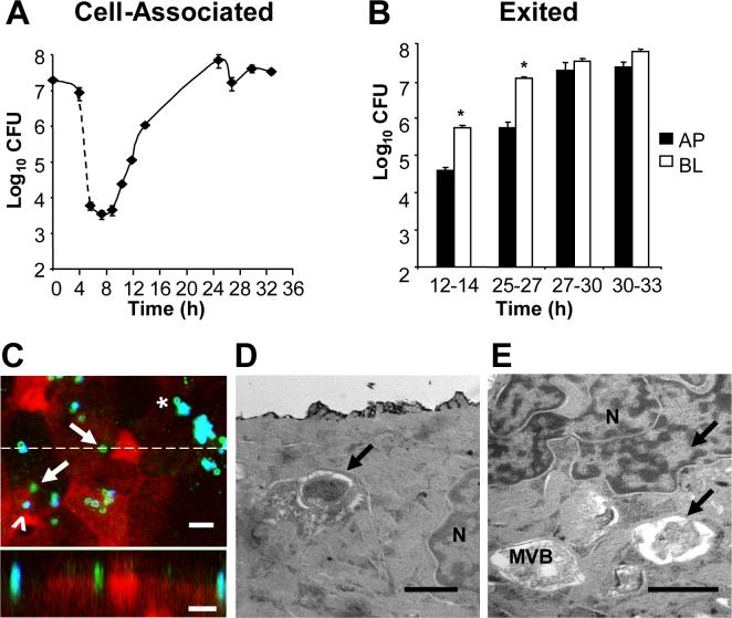
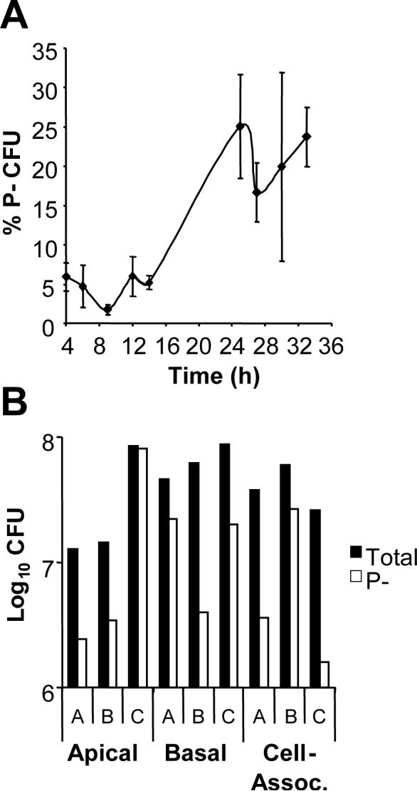
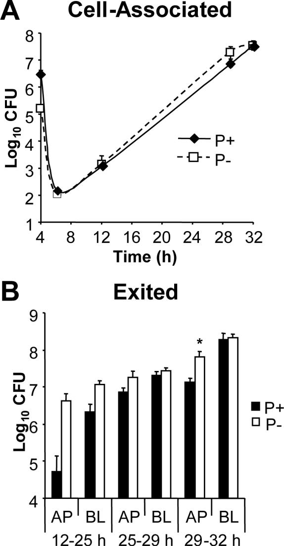
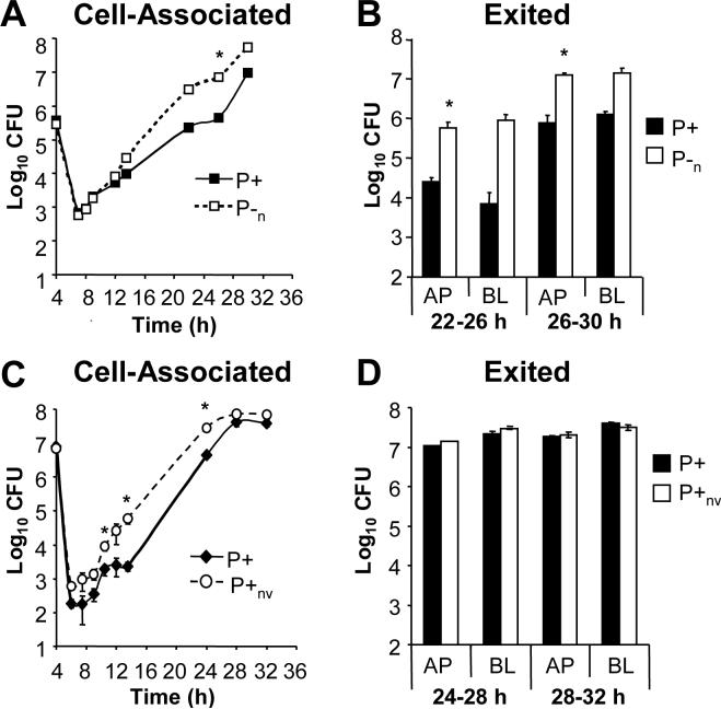
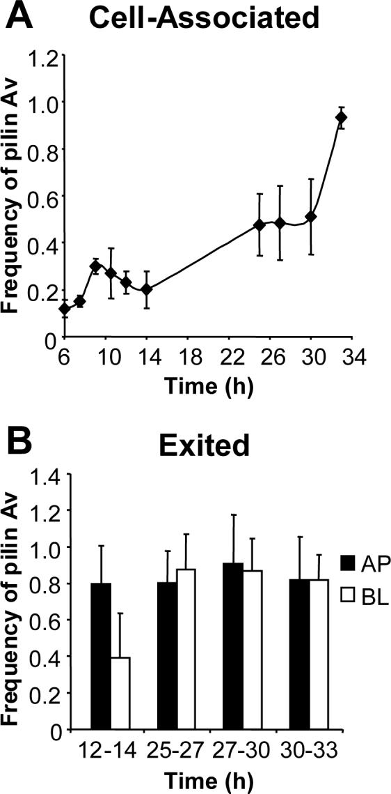
Type IV pili are a major virulence factor of the obligate human pathogen Neisseria gonorrhoeae (the gonococcus; Gc). Pili facilitate bacterial adherence to epithelial cells, but their participation in later steps of epithelial infection, particularly intracellular replication and exit, is poorly understood. Using polarized T84 cells as a model for mature mucosal epithelia, pilus dynamics in piliated, Opa-expressing Gc were examined over time. T84 infection was characterized by a several-hour delay in the growth of cell-associated bacteria and by non-directional exit of Gc, the first time these phenomena have been reported. During infection, non-piliated progeny arose stochastically from piliated progenitors. Piliated and non-piliated Gc replicated and exited from T84 cell monolayers equally well, demonstrating that piliation did not influence Gc survival during epithelial infection. The frequency with which pilin variants arose from a defined piliated progenitor during T84 cell infection was found to be sufficiently high to account for the extensive pilin variation reported during human infection. However, the repertoire of variants appearing in association with T84 cells was similar to what was seen in the absence of cells, demonstrating that polarized epithelial cells can support Gc replication without selecting for a subset of pilin variants or piliation states.


Figures

Figure 1
Figure 1. Profile of Gc infection of polarized T84 cells
Panel A: Cell-associated Gc. Polarized monolayers of 106 T84 cells were apically infected in triplicate with P+ FA1090 Gc for 4 h, then treated with gentamicin for 2 h (dotted line). At the indicated times, T84 lysates were serially diluted and plated for colony-forming units (CFU). The average number of CFU ± standard error of the mean (SEM) for three replicate monolayers per time point is presented. Panel B: Exited Gc. At the indicated intervals, the apical (AP; black bars) and basal (BL; white bars) medium was removed from infected T84 cells and CFU enumerated from each. *, P < 0.01 between apical and basal CFU in the specified interval (Student's t-test). Panel C: Gc are detected within T84 cells. T84 cells were fixed at 12 h post-infection and intracellular and extracellular Gc were discriminated from one another by differential antibody accessibility and examined by confocal laser scanning miscroscopy. Green, intracellular Gc; turquoise (blue + green), extracellular Gc; epithelial F-actin, red. In the top image, arrows point to two intracellular bacteria, one of which is adjacent to an extracellular bacterium (arrowhead). Asterisk denotes a potentially intracellular bacterium at the edge of a microcolony. In the bottom image, an X-Z section was taken through the slice indicated in the top image by the dotted line and shows an internalized, subapical bacterium in the middle of the field, flanked by two apically adherent, extracellular bacteria. Scale bars, 5 μm. Panels D-E: Gc reside within vacuoles in T84 cells. T84 cells were fixed at 12 h post-infection and processed for thin-section transmission electron microscopy. Arrows indicate intracellular bacteria; N, nucleus; MVB, multivesicular body. Scale bars, 1 μm.
Figure 2
Figure 2. P− Gc variants arise from a P+ progenitor during T84 cell infection
Panel A: Percentage of cell-associated P− CFU. The pilus-dependent colony morphology of CFU arising over time in T84 cells infected with P+ FA1090 Gc was examined using a stereomicroscope. The average percentage of P− CFU ± SEM was determined for three replicate monolayers per time point. Panel B: P− Gc arise stochastically during epithelial infection. Three monolayers of T84 cells, denoted A-C, were infected with P+ FA1090. The P+ (black bars) and P− (white bars) CFU present in the cell-associated population of each monolayer at 30 h and entering the apical and basal medium from each monolayer between 27 and 30 h were enumerated as in Figs. 1A and B.
Figure 3
Figure 3. Piliation is dispensable for Gc replication and exit from T84 cells
T84 cells were apically infected with P+ FA1090 Gc (solid lines, black bars) or an isogenic P− pilin variant (P−; dotted lines, white bars). Panel A: Cell-associated CFU. The CFU associated with the T84 cells (Panel A) were enumerated as in Fig. 1A. Panel B: Exited CFU. CFU exiting into the apical (AP) and basal (BL) medium (Panel B) were enumerated as in Fig. 1B. *, P < 0.05 by Student's t-test.
Figure 4
Figure 4. Gc that are nonvarying in P+ or P− phenotype can replicate and exit from T84 cells similar to the P+ parental strain
Panels A-B: Comparison of varying P+ to nonvarying P− FA1090 Gc. T84 cells were apically infected with the P+ FA1090 parental strain (solid lines; black bars) or an isogenic P−n mutant with a deletion in the pilE locus (dotted lines; white bars). The number of cell-associated (Panel A) and exited (Panel B) CFU were enumerated over time as in Figs. 1A and B, respectively. Panels C-D: Comparison of varying P+ to nonvarying P+ FA1090. T84 cells were apically infected with the P+ FA1090 parent (solid lines; black bars) or an isogenic P+nv mutant that does not undergo pilin Av due to the presence of a transposon insertion upstream of pilE (dotted lines; white bars). The number of cell-associated (Panel C) and exited (Panel D) CFU were enumerated over time as in Figs. 1A and B, respectively. *, P < 0.05 between strains at the indicated time point by Student's t-test.
Figure 5
Figure 5. Extensive pilin antigenic variation occurs in Gc associated with T84 cells
Panel A: Cell-associated CFU. T84 cells were infected with P+ FA1090, and CFU of varying pilus-dependent morphologies were collected from cell lysates. Panel B: Exited CFU. CFU were collected from the apical (AP; black bars) and basal (BL; white bars) medium of infected T84 cells at the indicated intervals. The pilE genes of the collected CFU were amplified, sequenced, and examined for changes from the parental 1−81-S2 sequence. The frequency of pilin Av at each time point or interval is defined as the number of recombination events detected divided by the number of pilE genes sequenced. The frequency was measured separately for the P+ and PCFU in each population, and the overall frequency of pilin Av was calculated by the formula: Frequency = (Frequency P+)(% P+ CFU) + (Frequency P−)(% P− CFU). Data are presented as the average frequency of pilin Av ± SEM measured in triplicate T84 cell monolayers. From the data in Table 1, the actual frequency of pilin Av at each time point is approximately 55% of the reported values.

Similar articles

Cited by

References

    1. Apicella MA, Ketterer M, Lee FK, Zhou D, Rice PA, Blake MS. The pathogenesis of gonococcal urethritis in men: confocal and immunoelectron microscopic analysis of urethral exudates from men infected with Neisseria gonorrhoeae. J Infect Dis. 1996;173:636–646. - PubMed
    1. Ayala P, Lin L, Hopper S, Fukuda M, So M. Infection of epithelial cells by pathogenic neisseriae reduces the levels of multiple lysosomal constituents. Infect Immun. 1998;66:5001–5007. - PMC - PubMed
    1. Bergstrom S, Robbins K, Koomey JM, Swanson J. Piliation control mechanisms in Neisseria gonorrhoeae. Proc Natl Acad Sci USA. 1986;83:3890–3894. - PMC - PubMed
    1. Binnicker MJ, Williams RD, Apicella MA. Infection of human urethral epithelium with Neisseria gonorrhoeae elicits an upregulation of host anti-apoptotic factors and protects cells from staurosporine-induced apoptosis. Cell Microbiol. 2003;5:549–560. - PubMed
    1. Biswas GD, Sox T, Blackman E, Sparling PF. Factors affecting genetic transformation of Neisseria gonorrhoeae. J Bacteriol. 1977;129:983–992. - PMC - PubMed

Publication types

MeSH terms

Substances

LinkOut - more resources