

Globally distributed mycobacterial fish pathogens produce a novel plasmid-encoded toxic macrolide, mycolactone F
- PMID: 16923788
- PMCID: PMC1695495
- DOI: 10.1128/IAI.00970-06
Globally distributed mycobacterial fish pathogens produce a novel plasmid-encoded toxic macrolide, mycolactone F
Abstract
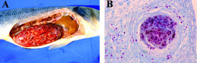
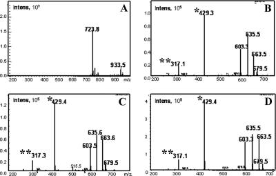
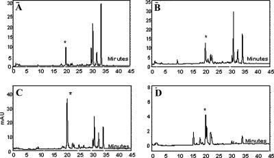
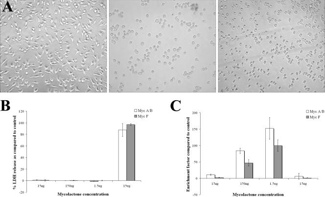
Mycobacterium ulcerans and Mycobacterium marinum are closely related pathogens which share an aquatic environment. The pathogenesis of these organisms in humans is limited by their inability to grow above 35 degrees C. M. marinum causes systemic disease in fish but produces localized skin infections in humans. M. ulcerans causes Buruli ulcer, a severe human skin lesion. At the molecular level, M. ulcerans is distinguished from M. marinum by the presence of a virulence plasmid which encodes a macrolide toxin, mycolactone, as well as by hundreds of insertion sequences, particularly IS2404. There has been a global increase in reports of fish mycobacteriosis. An unusual clade of M. marinum has been reported from fish in the Red and Mediterranean Seas and a new mycobacterial species, Mycobacterium pseudoshottsii, has been cultured from fish in the Chesapeake Bay, United States. We have discovered that both groups of fish pathogens produce a unique mycolactone toxin, mycolactone F. Mycolactone F is the smallest mycolactone (molecular weight, 700) yet identified. The core lactone structure of mycolactone F is identical to that of M. ulcerans mycolactones, but a unique side chain structure is present. Mycolactone F produces apoptosis and necrosis on cultured cells but is less potent than M. ulcerans mycolactones. Both groups of fish pathogens contain IS2404. In contrast to M. ulcerans and conventional M. marinum, mycolactone F-producing mycobacteria are incapable of growth at above 30 degrees C. This fact is likely to limit their virulence for humans. However, such isolates may provide a reservoir for horizontal transfer of the mycolactone plasmid in aquatic environments.
Figures

Similar articles
-
A newly discovered mycobacterial pathogen isolated from laboratory colonies of Xenopus species with lethal infections produces a novel form of mycolactone, the Mycobacterium ulcerans macrolide toxin.Infect Immun. 2005 Jun;73(6):3307-12. doi: 10.1128/IAI.73.6.3307-3312.2005. Infect Immun. 2005. PMID: 15908356 Free PMC article.
-
Mycobacterium ulcerans toxic macrolide, mycolactone modulates the host immune response and cellular location of M. ulcerans in vitro and in vivo.Cell Microbiol. 2005 Sep;7(9):1295-304. doi: 10.1111/j.1462-5822.2005.00557.x. Cell Microbiol. 2005. PMID: 16098217
-
Evolution of Mycobacterium ulcerans and other mycolactone-producing mycobacteria from a common Mycobacterium marinum progenitor.J Bacteriol. 2007 Mar;189(5):2021-9. doi: 10.1128/JB.01442-06. Epub 2006 Dec 15. J Bacteriol. 2007. PMID: 17172337 Free PMC article.
-
Mycolactone: More than Just a Cytotoxin.2019 Apr 30. In: Pluschke G, Röltgen K, editors. Buruli Ulcer: Mycobacterium Ulcerans Disease [Internet]. Cham (CH): Springer; 2019. 2019 Apr 30. In: Pluschke G, Röltgen K, editors. Buruli Ulcer: Mycobacterium Ulcerans Disease [Internet]. Cham (CH): Springer; 2019. PMID: 32091711 Free Books & Documents. Review.
-
Pathogenetic mechanisms of the intracellular parasite Mycobacterium ulcerans leading to Buruli ulcer.Lancet Infect Dis. 2009 Nov;9(11):699-710. doi: 10.1016/S1473-3099(09)70234-8. Lancet Infect Dis. 2009. PMID: 19850228 Review.
Cited by
-
Mycobacterium pseudoshottsii in Mediterranean Fish Farms: New Trouble for European Aquaculture?Pathogens. 2020 Jul 27;9(8):610. doi: 10.3390/pathogens9080610. Pathogens. 2020. PMID: 32726963 Free PMC article.
-
Deciphering the genetic basis for polyketide variation among mycobacteria producing mycolactones.BMC Genomics. 2008 Oct 7;9:462. doi: 10.1186/1471-2164-9-462. BMC Genomics. 2008. PMID: 18840298 Free PMC article.
-
Total Synthesis of Mycolactones A and B.Tetrahedron. 2007 Jun 25;63(26):5739-5753. doi: 10.1016/j.tet.2007.02.057. Tetrahedron. 2007. PMID: 17940589 Free PMC article.
-
Complete Genome and Partial Megaplasmid Sequences of Mycobacterium pseudoshottsii Strain NJB1907-Z4, Isolated from an Aquarium-Reared Japanese Sardine (Sardinops melanostictus) in Japan.Microbiol Resour Announc. 2022 Dec 15;11(12):e0078522. doi: 10.1128/mra.00785-22. Epub 2022 Nov 9. Microbiol Resour Announc. 2022. PMID: 36350130 Free PMC article.
-
Mycolactone gene expression is controlled by strong SigA-like promoters with utility in studies of Mycobacterium ulcerans and buruli ulcer.PLoS Negl Trop Dis. 2009 Nov 24;3(11):e553. doi: 10.1371/journal.pntd.0000553. PLoS Negl Trop Dis. 2009. PMID: 19936295 Free PMC article.
References
-
- Adusumilli, S., A. Mve-Obiang, T. Sparer, W. Meyers, J. Hayman, and P. L. C. Small. 2005. Mycobacterium ulcerans toxic macrolide, mycolactone modulates the host immune response and cellular location of M. ulcerans in vitro and in vivo. Cell. Microbiol. 7:1295-1304. - PubMed
-
- Asiedu, K., R. Scherpbier, and M. Raviglione (ed.). 2000. Buruli ulcer: Mycobacterium ulcerans infection. World Health Organization, Geneva, Switzerland.
-
- Barker, D. J. 1973. Epidemiology of Mycobacterium ulcerans infection. Trans. R. Soc. Trop. Med. Hyg. 67:43-50. - PubMed
-
- Cadapan, L. D., R. L. Arslanian, J. R. Carney, S. M. Zavala, P. L. Small, and P. Licari. 2001. Suspension cultivation of Mycobacterium ulcerans for the production of mycolactones. FEMS Microbiol. Lett. 10246:1-5. - PubMed
-
- Colorni, A. 1992. A systemic mycobacteriosis in the European sea bass Dicentrarchus labrax cultured in Eilat (Red Sea). Isr. J. Aquacult. Bamidgeh. 44:75-81.
Publication types
MeSH terms
Substances
Associated data
- Actions
- Actions
- Actions
- Actions
- Actions
- Actions
Grants and funding
LinkOut - more resources
Full Text Sources
Medical
Molecular Biology Databases
