Comparative Study
. 2006 Nov;74(11):6365-76.
doi: 10.1128/IAI.00974-06. Epub 2006 Aug 21.

Discordance in the effects of Yersinia pestis on the dendritic cell functions manifested by induction of maturation and paralysis of migration

Affiliations
Comparative Study

Discordance in the effects of Yersinia pestis on the dendritic cell functions manifested by induction of maturation and paralysis of migration

Baruch Velan et al. Infect Immun. 2006 Nov.

Abstract

The encounter between invading microorganisms and dendritic cells (DC) triggers a series of events which include uptake and degradation of the microorganism, induction of a maturation process, and enhancement of DC migration to the draining lymph nodes. Various pathogens have developed strategies to counteract these events as a measure to evade the host defense. In the present study we found that interaction of the Yersinia pestis EV76 strain with DC has no effect on cell viability and is characterized by compliance with effective maturation, which is manifested by surface display of major histocompatibility complex class II, of costimulatory markers, and of the chemokine receptor CCR7. This is in contrast to maturation inhibition and cell death induction exerted by the related species Yersinia enterocolitica WA O:8. Y. pestis interactions with DC were found, however, to impair functions related to cytoskeleton rearrangement. DC pulsed with Y. pestis failed to adhere to solid surfaces and to migrate toward the chemokine CCL19 in an in vitro transmembrane assay. Both effects were dependent on the presence of the pCD1 virulence plasmid and on a bacterial growth shift to 37 degrees C prior to infection. Moreover, while instillation of a pCD1-cured Y. pestis strain into mouse airways triggered effective transport of alveolar DC to the mediastinal lymph node, instillation of Y. pestis harboring the plasmid failed to do so. Taken together, these results suggest that virulence plasmid-dependent impairment of DC migration is the major mechanism utilized by Y. pestis to subvert DC function.


Figures

FIG. 1.
FIG. 1.
Effect of Yersinia infection on DC viability. BMDC were pulsed at an MOI of 20 with the indicated bacterial strain by centrifugation. Gentamicin (20 μg/ml) was added 1 h later, and incubation was carried out for 4, 12, or 24 h. Cells were then stained with annexin V conjugated to Alexa 568 and propidium iodide and analyzed by flow cytometry. (A) Cell death at 12 h postinfection is expressed in the form of dot plots, and numbers adjacent to plots indicate the percentage of cells in each quadrant. (B) Summary of cell death data at 4, 12, and 24 h postinfection, presented as bar diagrams depicting the ratio between the various quadrants (UR, annexin+/PI+; LR, annexin/PI+; UL, annexin+/PI; LL, annexin/PI). Results are representative of three independent experiments.
FIG. 2.
FIG. 2.
Expression of costimulatory molecule and MHC class II on DC infected with Y. pestis. Cells were pulsed by centrifugation with LPS or with bacteria (shifted to grow at 37°C) at an MOI of 50. One hour later, 20 μg/ml gentamicin was added and cells were incubated for 23 h. Staining for the indicated surface markers and flow cytometry were performed as described in Materials and Methods. Gray areas denote the isotype-matched immunoglobulin control. Mean fluorescence intensities for surface marker staining are given at the right top corner of each diagram. Mean fluorescence intensities for isotype controls in all experiments presented in this figure ranged between 3 (class II) and 50 (CD83) and are therefore not presented. Three independent experiments exhibited similar results. Very similar results were obtained with an alternative infection protocol in which bacteria were washed away at 1 h after pulsing and cells were incubated in gentamicin-containing fresh medium for 23 h. In all these experiments optimal activation was observed at an MOI of 50, yet the same pattern of up-regulation was also obtained at an MOI of 20.
FIG. 3.
FIG. 3.
Expression of CD54 on DC infected with Y. pestis or Y. enterocolitica strains. Cells were pulsed with the indicated bacterial strains as described in the legend to Fig. 2, except that incubation was terminated at 4 h postinfection. Mean fluorescence intensities for surface marker staining are given at the right top corner of each diagram. Mean fluorescence intensities for isotype controls ranged from 4.5 to 5.5. Three independent experiments exhibited similar results.
FIG. 4.
FIG. 4.
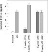
Secretion of TNF-α by DC infected with Y. pestis. Cells were pulsed with the indicated bacterial strains at an MOI of 5 (bacteria were grown for 2.5 h at the indicated temperature prior to infection) or with LPS. One hour later, 20 μg/ml gentamicin was added and cells were incubated for 23 h. TNF-α was quantified in cell medium as described in Materials and Methods. Each experiment was performed in triplicate, and data represent averages and standard deviations. Two additional experiments performed under similar conditions and one experiment conducted at an MOI of 20 exhibited similar results.
FIG. 5.
FIG. 5.
Effect of Y. pestis infection on DC transmembrane migration. Cells were pulsed without centrifugation with the indicated bacterial strains at an MOI of 5 (higher MOIs had adverse effects on DC migration independent of the Yersinia strain used). Gentamicin was added at 0.5 h after pulsing for infection with bacteria grown at 28°C and at 1 h for bacteria grown at 37°C. Twenty-four hours after pulsing, cells were examined for migration in Transwell chambers towards CCL19 (A) or control medium (B). Results are presented as percent cells migrating from the upper compartments to the lower compartment. Each experiment was performed in triplicate, and data represent averages and standard deviations. Two additional experiments performed under similar conditions exhibited similar results.
FIG. 6.
FIG. 6.
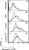
Expression of CCR7 on DC infected with Y. pestis strains. Cells were infected and analyzed for CCR7 expression as described in legend to Fig. 3. Gray lines denote the isotype-matched immunoglobulin control. Mean fluorescence intensities for surface marker staining are given at the right top corner of each diagram. Mean fluorescence intensities for isotype controls are given at left top corner. Results are representatives of four independent experiments.
FIG. 7.
FIG. 7.
Effect of Y. pestis infection on DC adhesion. Pulsing was performed in 96-well microtiter plates at an MOI of 50 by centrifugation, and gentamicin (20 μg/ml) was added 1 h later. At the indicated times, medium and nonadherent cells were washed away, and the quantity of adhered cells was monitored by determining their LDH content (see Materials and Methods). Data are presented as the percentage of bound cells of those applied to the well (LDH content in bound cells of LDH content in input). (A) Comparison of adhesion induced at 1.5 h after pulsing with Y. pestis strains grown at 37°C to that induced by LPS and to spontaneous adhesion. (B) Kinetics of adhesion after pulsing with Y. pestis (black squares) and Y. pestis Δ70 (hatched squares) grown at 37°C (upper panel) and 28°C lower panel). Data are from a typical representative of three experiments, each performed in triplicates. Error bars indicate standard deviations.
FIG. 8.
FIG. 8.
Migration of respiratory DC to draining lymph nodes after intranasal instillation of Y. pestis. Y. pestis and Y. pestis Δ70 suspensions were administered by intranasal instillation (two mice per group) in murine respiratory tracts prestained with CMTMR (see Materials and Methods). Instillation of 30 μg of LPS or PBS alone served as controls. After 18 h, mediastinal lymph nodes were examined by flow cytometry for evidence of DC immigrating from the airways. Two regions, R1 (CMTMR+/CD11c high) and R2 (CMTMR+/CD11cmedium), were gated. The tables adjacent to the dot plots provide gate statistics for staining with anti-CD11c and with the isotype-matched immunoglobulin control (isoC). Dot plots for isotype control staining are not shown. Two additional experiments performed under similar conditions exhibited similar results.

Similar articles

Cited by

References

    1. Banchereau, J., and R. M. Steinman. 1998. Dendritic cells and the control of immunity. Nature 392:245-252. - PubMed
    1. Ben-Gurion, R., and A. Shafferman. 1981. Essential virulence determinants of different Yersinia species are carried on a common plasmid. Plasmid 5:183-187. - PubMed
    1. Ber, R., E. Mamroud, M. Aftalion, A. Tidhar, D. Gur, Y. Flashner, and S. Cohen. 2003. Development of an improved selective agar medium for isolation of Yersinia pestis. Appl. Environ. Microbiol. 69:5787-5792. - PMC - PubMed
    1. Billard, E., C. Cazevieille, J. Dornand, and A. Gross. 2005. High susceptibility of human dendritic cells to invasion by the intracellular pathogens Brucella suis, B. abortus, and B. melitensis. Infect. Immun. 73:8418-8424. - PMC - PubMed
    1. Boland, A., and G. R. Cornelis. 1998. Role of YopP in suppression of tumor necrosis factor alpha release by macrophages during Yersinia infection. Infect. Immun. 66:1878-1884. - PMC - PubMed

Publication types

MeSH terms

LinkOut - more resources