. 2006 Sep 4;203(9):2165-75.
doi: 10.1084/jem.20061022. Epub 2006 Aug 21.

Complement activation induces dysregulation of angiogenic factors and causes fetal rejection and growth restriction

Affiliations

Complement activation induces dysregulation of angiogenic factors and causes fetal rejection and growth restriction

Guillermina Girardi et al. J Exp Med. .

Abstract

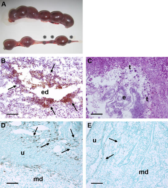
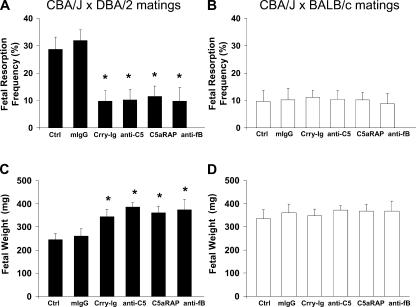
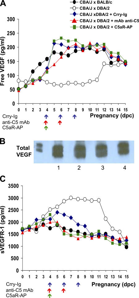
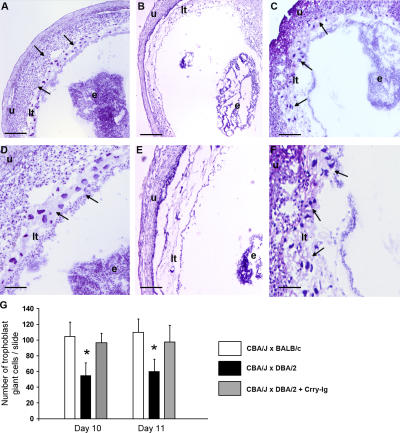
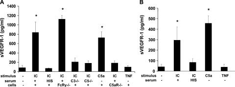
Immune mechanisms have been implicated in placental dysfunction in patients with recurrent miscarriages and intrauterine growth restriction (IUGR), but the mediators are undefined. Here we show that complement activation, particularly C5a, is a required intermediary event in the pathogenesis of placental and fetal injury in an antibody-independent mouse model of spontaneous miscarriage and IUGR, and that complement activation causes dysregulation of the angiogenic factors required for normal placental development. Pregnancies complicated by miscarriage or growth restriction were characterized by inflammatory infiltrates in placentas, functional deficiency of free vascular endothelial growth factor (VEGF), elevated levels of soluble VEGF receptor 1 (sVEGFR-1, also known as sFlt-1; a potent anti-angiogenic molecule), and defective placental development. Inhibition of complement activation in vivo blocked the increase in sVEGFR-1 and rescued pregnancies. In vitro stimulation of monocytes with products of the complement cascade directly triggered release of sVEGFR-1, which sequesters VEGF. These studies provide the first evidence linking the complement system to angiogenic factor imbalance associated with placental dysfunction, and identify a new effector of immune-triggered pregnancy complications.


Figures

Figure 1.
Figure 1.
Resorption of embryos, deposition of complement C3, and infiltration of monocytes in decidual tissue of DBA/2-mated CBA mice. (A) Representative uteri from mice killed at day 15 of pregnancy are shown. The top panel, from a BALB/c-mated CBA/J mouse contains larger amnion sacs and no resorptions. The bottom panel, from a DBA/2-mated CBA/J mouse, shows six amnion sacs of varying sizes and three resorptions (asterisks). (B–E) Pregnant mice were killed on day 8, and sections were stained with anti–mouse C3 (B and C) or anti–mouse F4/80 (D and E) to detect monocytes. In CBA/J × DBA/2 mice, there was extensive C3 deposition (brown, arrows; B) and monocyte infiltration (arrows) in the deciduas (D). In contrast, the decidual tissue from CBA/J × BALB/c mice showed minimal staining for C3 (C) and monocyte infiltration (E). e, embryo; ed, embryonic debris; md, maternal deciduas; t, trophoblasts; u, uterine wall. Bars, 0.1 mm.
Figure 2.
Figure 2.
Inhibition of complement prevents fetal death and growth restriction in CBA/J × DBA/2 matings. DBA/2- and BALB/c-mated CBA/J females were treated with recombinant Crry-Ig, anti-C5 mAb (mAb BB5.1), C5aR antagonist peptide (C5aRAP) (AcPhe[l-ornithine-Pro-d-cyclohexylalanine-Trp-Arg]), anti–factor B mAb (mAb 1379), or mouse polyclonal IgG (mIgG). Mice were killed on day 15 of pregnancy, uteri were dissected, embryos were weighed, and fetal resorption rates were calculated (number of resorptions/number of fetuses + number of resorptions). There were six to eight mice in each experimental group. (A and B) The frequency of fetal resorptions in treated and untreated CBA/J × DBA/2 and CBA/J × BALB/c pregnancies was determined (CBA/J × DBA/2 vs. CBA/J × BALB/c, P < 0.001; CBA/J × DBA/2 vs. CBA/J × DBA/2 plus complement inhibitors; *, P < 0.001). (C and D) Average fetal weights in treated and untreated CBA/J × DBA/2 and CBA/J × BALB/c pregnancies was determined (CBA/J × DBA/2 vs. CBA/J × BALB/c, P < 0.005; CBA/J × DBA/2 vs. CBA/J ×DBA/2 plus complement inhibitors; *, P < 0.005).
Figure 3.
Figure 3.
Dysregulation of angiogenic factors in CBA/J × DBA/2 matings is prevented by inhibiting complement. Free VEGF, total VEGF, and sVEGFR-1 were assessed. Free VEGF (A) and total sVEGFR-1 (C) levels in plasma were measured in CBA/J × DBA/2 (◯) and CBA/J × BALB/c (•) mice from days 1 through 15 of pregnancy. Some DBA/2-mated CBA/J mice were treated with Crry-IgG (♦), anti-C5 mAb (▴), and C5aR-AP (▪). The arrows indicate the days when the complement inhibitors were administered. Values represent means from three to five mice/time point/group (free VEGF: CBA/J × DBA/2 vs. CBA/J × BALB/c, P < 0.001; CBA/J × DBA/2 vs. CBA/J × DBA/2 plus complement inhibitors, P < 0.001; sVEGFR-1: CBA/J × DBA/2 vs. CBA/J × BALB/c, P < 0.001; CBA/J × DBA/2 vs. CBA/J × DBA/2 plus complement inhibitors, P < 0.005). (B) Total VEGF levels were compared in CBA/J × BALB/c and CBA/J × DBA/2 matings. Plasma samples from early pregnancy (days 5–8) and late pregnancy (days 9–12) were pooled into two groups for detection of total plasma VEGF (bound to sVEGFR-1 and unbound) by Western blotting. Samples were immunoprecipitated with rabbit anti–mouse VEGF and analyzed with anti-VEGF mAb. Lane 1, CBA/J × BALB/c days 5–8; lane 2, CBA/J × BALB/c days 9–12; lane 3, CBA/J × DBA/2 days 5–8; lane 4, CBA/J × DBA/2 days 9–12.
Figure 4.
Figure 4.
Decreased trophoblast giant cells in placentas from CBA/J × DBA/2 matings. (A–F) Histologic analysis of sections of uteri from CBA/J × BALB/c matings (A and D), CBA/J × DBA/2 (B and E) matings, and (C and F) CBA/J × DBA/2 matings treated with Crry-Ig killed at day 10 of pregnancy. Trophoblast giant cells (arrows) were reduced in midtrimester placentas from DBA/2-mated CBA/J pregnancies (B and E) compared with CBA/2 × BALB/c pregnancies, and this was prevented by treatment with Crry-Ig. Sections were stained with hematoxylin and eosin. e, embryo; lt, labyrinthine trophoblast u, uterine wall. Scale bars, 0.1 mm (A–C); 0.025 mm (D–F). (G) Number of trophoblast giant cells in placentas from days 10 and 11 of pregnancy was counted by light microscopy. Data are expressed as the mean of four sections counted by two readers blinded to experimental conditions. *, P < 0.01.
Figure 5.
Figure 5.
Complement activation triggers release of sVEGFR-1 by murine macrophages and human monocytes in vitro. (A) Splenic macrophages from CBA/J female mice were stimulated with heat-aggregated human IgG (IC) in the presence of 10% normal mouse serum, 10% heat-inactivated mouse serum (HIS), 10% serum from C3-deficient mice (C3−/−), or 10% serum from C5-deficient mice (C5−/−). Macrophages were also incubated with C5a, TNF-α, or control medium. Splenic macrophages from mice deficient in stimulatory Fcγ receptors (FcRγ−/−) or C5aR (C5aR−/−) were also treated with ICs. Culture supernatants were collected after 24 h and analyzed for sVEGFR-1 by ELISA (n = 3–9 experiments/group; *, P < 0.001 vs. control). (B) Adherent human mononuclear cells from healthy donors were stimulated with ICs plus 10% normal human serum and ICs plus 10% heat-inactivated human serum, C5a, or TNF-α. After 2 h, culture supernatants were collected and analyzed for sVEGFR-1 by ELISA (n = 4–5 experiments/group; *, P < 0.005 vs. control).

References

    1. Mills, J.L., J.L. Simpson, S.G. Driscoll, L. Jovanovic-Peterson, M. Van Allen, J.H. Aarons, B. Metzger, F.R. Bieber, R.H. Knopp, L.B. Holmes, et al. 1988. Incidence of spontaneous abortion among normal women and insulin-dependent diabetic women whose pregnancies were identified within 21 days of conception. N. Engl. J. Med. 319:1617–1623. - PubMed
    1. Resnik, R., and R.K. Creasy. 2003. Intrauterine growth restriction. In Maternal-Fetal Medicine: Principles and Practice. J.D. Iams, editor. Saunders, Philadelphia. 495–512.
    1. Godfrey, K.M., and D.J. Barker. 2000. Fetal nutrition and adult disease. Am. J. Clin. Nutr. 71:1344S–1352S. - PubMed
    1. Mellor, A.L., and D.H. Munn. 2000. Immunology at the maternal-fetal interface: lessons for T cell tolerance and suppression. Annu. Rev. Immunol. 18:367–391. - PubMed
    1. Hunt, J.S., M.G. Petroff, R.H. McIntire, and C. Ober. 2005. HLA-G and immune tolerance in pregnancy. FASEB J. 19:681–693. - PubMed

Publication types

MeSH terms

Substances