. 2006 Sep;188(17):6081-91.
doi: 10.1128/JB.00338-06.

The structure of MbtI from Mycobacterium tuberculosis, the first enzyme in the biosynthesis of the siderophore mycobactin, reveals it to be a salicylate synthase

Affiliations

The structure of MbtI from Mycobacterium tuberculosis, the first enzyme in the biosynthesis of the siderophore mycobactin, reveals it to be a salicylate synthase

Anthony J Harrison et al. J Bacteriol. 2006 Sep.

Abstract

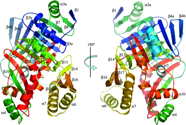
The ability to acquire iron from the extracellular environment is a key determinant of pathogenicity in mycobacteria. Mycobacterium tuberculosis acquires iron exclusively via the siderophore mycobactin T, the biosynthesis of which depends on the production of salicylate from chorismate. Salicylate production in other bacteria is either a two-step process involving an isochorismate synthase (chorismate isomerase) and a pyruvate lyase, as observed for Pseudomonas aeruginosa, or a single-step conversion catalyzed by a salicylate synthase, as with Yersinia enterocolitica. Here we present the structure of the enzyme MbtI (Rv2386c) from M. tuberculosis, solved by multiwavelength anomalous diffraction at a resolution of 1.8 A, and biochemical evidence that it is the salicylate synthase necessary for mycobactin biosynthesis. The enzyme is critically dependent on Mg2+ for activity and produces salicylate via an isochorismate intermediate. MbtI is structurally similar to salicylate synthase (Irp9) from Y. enterocolitica and the large subunit of anthranilate synthase (TrpE) and shares the overall architecture of other chorismate-utilizing enzymes, such as the related aminodeoxychorismate synthase PabB. Like Irp9, but unlike TrpE or PabB, MbtI is neither regulated by nor structurally stabilized by bound tryptophan. The structure of MbtI is the starting point for the design of inhibitors of siderophore biosynthesis, which may make useful lead compounds for the production of new antituberculosis drugs, given the strong dependence of pathogenesis on iron acquisition in M. tuberculosis.


Figures

FIG. 1.
FIG. 1.
Chemical structure of mycobactin T, the siderophore of M. tuberculosis, annotated with the biosynthetic origins of its constituent components. In water-soluble variants, the acyl chain on the central modified lysine is 2 to 8 carbons long; in cell-wall associated variants, it can be up to 20 carbons in length. The drawing is based on data from reference .
FIG. 2.
FIG. 2.
Structurally characterized chorismate-utilizing pathways. Shown are the three analogous transformations of chorismate for which there is structural information about the enzymes involved: anthranilate synthesis, p-aminobenzoate synthesis, and salicylate synthesis. ADC, 4-amino4-deoxychorismate; ADIC, 2-amino-2-deoxyisochorismate.
FIG. 3.
FIG. 3.
Ribbon diagram of the monomer structure of MbtI, colored blue to red from the N to the C terminus, with secondary structure elements as defined by DSSP, and labeled to be consistent with the nomenclature used for TrpE (50). The active site cleft is highlighted with a cyan circle.
FIG. 4.
FIG. 4.
Structure-based multiple sequence alignment of MbtI with related enzymes of known structure. Irp9, salicylate synthase from Y. enterocolitica (PDB accession no. 2FN0/2FN1); PabB, p-aminobenzoate synthase from E. coli (PDB accession no. 1K0E/1K0G); TrpE, anthranilate synthase from S. marcescens (PDB accession no. 1I7Q/1I7S). Structural alignment was made using sPDBv (25) and subsequently edited by hand. The figure was drawn using ESPript (23). Identical residues are shown in white type with a black background, and similar residues which are conserved in all four sequences are shown boxed. (Similar groups as defined by ESPript are as follows: HKR, polar positive; DE, polar negative; STNQ, polar neutral; AVLIM, nonpolar aliphatic; FYW, nonpolar aromatic; PG, structure breakers; and C, thiol.) Secondary structure elements for MbtI are shown above the alignment, labeled to be consistent with the nomenclature used for TrpE (31, 50).
FIG. 5.
FIG. 5.
(A) Comparison of the active site regions of MbtI and the ligand-bound conformation of TrpE, showing the shift in the β14-β17-β16 sheet. MbtI is shown in green, and TrpE is shown in blue with side chains in yellow. The bound Mg2+ ion is shown as a yellow sphere. In all cases except where noted, molecule B from the MbtI asymmetric unit is shown. (B) Overlay of the MbtI, TrpE, and Irp9 active sites. MbtI is shown in green, TrpE in yellow, and Irp9 in blue. (C) Pyruvate binding in the active site of MbtI. A molecule of pyruvate is shown modeled into residual density from a SigmaA-weighted FoFc electron density map, contoured at 3σ, in the active site of molecule B in the asymmetric unit. Side chains are shown in green for residues that interact with the pyruvate, and hydrogen bonds are shown as dashed lines. This arrangement is identical in molecule A of the asymmetric unit. The alternative conformation of Arg405 in molecules C and D in the asymmetric unit, where no bound pyruvate is observed, is shown in light blue, represented using the side chain from molecule C. (D) The tryptophan binding site of TrpE is overlaid on the equivalent region of MbtI. The hydrogen bonding network in MbtI is shown as black dashed lines. Molecules are colored as described for panel A.
FIG. 6.
FIG. 6.
Enzymatic activity of MbtI. (A) MbtI activity was monitored using fluorometric detection at an emission wavelength of 410 nm in the presence (+) and absence (−) of Mg2+. (B) Extracted ion chromatograms of MbtI reaction products, sampled over an m/z range from 137.0 to 137.1, compared to chromatograms of chorismate and salicylate incubated under the same conditions without enzyme. Chromatograms are all normalized to the highest peak. Chorismate yields a fragment with an m/z of 137 due to in-source fragmentation and also yields other fragments at an m/z of 207 and an m/z of 225 (traces not shown for clarity). (C) 1H-NMR spectroscopic analysis of salicylate production by MbtI, compared to spectra of chorismate and salicylate alone.

References

    1. Bauerle, R., J. Hess, and S. French. 1987. Anthranilate synthase-anthranilate phosphoribosyltransferase complex and subunits of Salmonella typhimurium. Methods Enzymol. 142:366-386. - PubMed
    1. Brunger, A. T., P. D. Adams, G. M. Clore, W. L. DeLano, P. Gros, R. W. Grosse-Kunstleve, J. S. Jiang, J. Kuszewski, M. Nilges, N. S. Pannu, R. J. Read, L. M. Rice, T. Simonson, and G. L. Warren. 1998. Crystallography & NMR system: a new software suite for macromolecular structure determination. Acta Crystallogr. D 54:905-921. - PubMed
    1. Bulloch, E. M. M., and C. Abell. 2005. Detection of covalent intermediates formed in the reaction of 4-amino-4-deoxychorismate synthase. Chembiochem 6:832-834. - PubMed
    1. Bulloch, E. M. M., M. A. Jones, E. J. Parker, A. P. Osborne, E. Stephens, G. M. Davies, J. R. Coggins, and C. Abell. 2004. Identification of 4-amino-4-deoxychorismate synthase as the molecular target for the antimicrobial action of (6S)-6-fluoroshikimate. J. Am. Chem. Soc. 126:9912-9913. - PubMed
    1. Clemens, D. L., and M. A. Horwitz. 1996. The Mycobacterium tuberculosis phagosome interacts with early endosomes and is accessible to exogenously administered transferrin. J. Exp. Med. 184:1349-1355. - PMC - PubMed

Publication types

Associated data

LinkOut - more resources