

Genome sequence and global gene expression of Q54, a new phage species linking the 936 and c2 phage species of Lactococcus lactis
- PMID: 16923877
- PMCID: PMC1595367
- DOI: 10.1128/JB.00581-06
Genome sequence and global gene expression of Q54, a new phage species linking the 936 and c2 phage species of Lactococcus lactis
Abstract
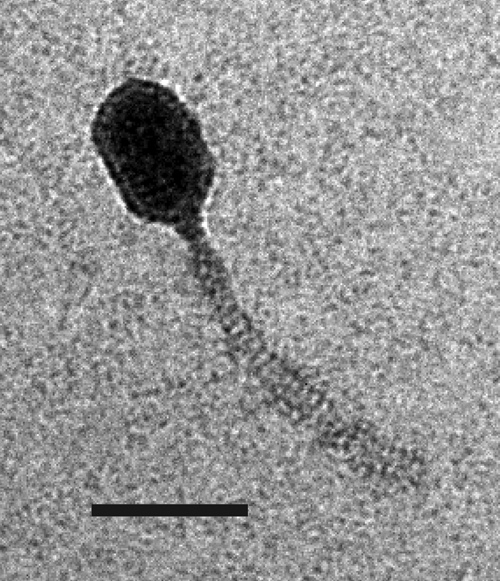
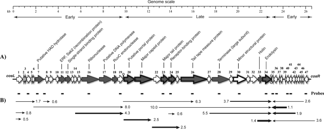
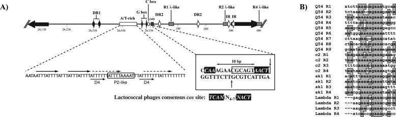
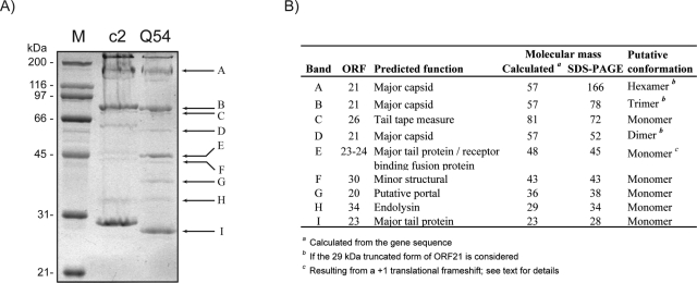
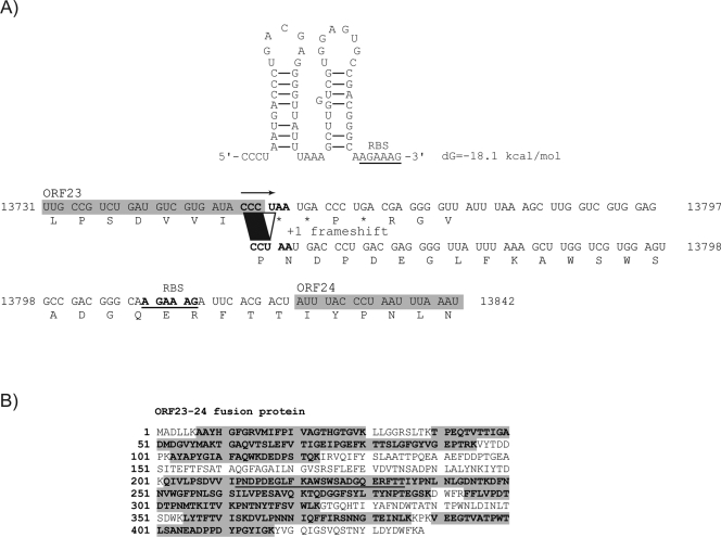
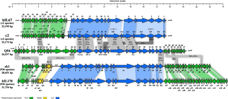
The lytic lactococcal phage Q54 was previously isolated from a failed sour cream production. Its complete genomic sequence (26,537 bp) is reported here, and the analysis indicated that it represents a new Lactococcus lactis phage species. A striking feature of phage Q54 is the low level of similarity of its proteome (47 open reading frames) with proteins in databases. A global gene expression study confirmed the presence of two early gene modules in Q54. The unusual configuration of these modules, combined with results of comparative analysis with other lactococcal phage genomes, suggests that one of these modules was acquired through recombination events between c2- and 936-like phages. Proteolytic cleavage and cross-linking of the major capsid protein were demonstrated through structural protein analyses. A programmed translational frameshift between the major tail protein (MTP) and the receptor-binding protein (RBP) was also discovered. A "shifty stop" signal followed by putative secondary structures is likely involved in frameshifting. To our knowledge, this is only the second report of translational frameshifting (+1) in double-stranded DNA bacteriophages and the first case of translational coupling between an MTP and an RBP. Thus, phage Q54 represents a fascinating member of a new species with unusual characteristics that brings new insights into lactococcal phage evolution.
Figures
Similar articles
-
A virulent phage infecting Lactococcus garvieae, with homology to Lactococcus lactis phages.Appl Environ Microbiol. 2015 Dec;81(24):8358-65. doi: 10.1128/AEM.02603-15. Epub 2015 Sep 25. Appl Environ Microbiol. 2015. PMID: 26407890 Free PMC article.
-
Genome and proteome of Listeria monocytogenes phage PSA: an unusual case for programmed + 1 translational frameshifting in structural protein synthesis.Mol Microbiol. 2003 Oct;50(1):303-17. doi: 10.1046/j.1365-2958.2003.03684.x. Mol Microbiol. 2003. PMID: 14507382
-
Genome analysis of the obligately lytic bacteriophage 4268 of Lactococcus lactis provides insight into its adaptable nature.Gene. 2006 Jan 17;366(1):189-99. doi: 10.1016/j.gene.2005.09.022. Epub 2005 Dec 1. Gene. 2006. PMID: 16325353
-
Species and type phages of lactococcal bacteriophages.Intervirology. 1991;32(1):2-9. doi: 10.1159/000150179. Intervirology. 1991. PMID: 1901837 Review.
-
Engineering of receptor-binding proteins in bacteriophages and phage tail-like bacteriocins.Biochem Soc Trans. 2019 Feb 28;47(1):449-460. doi: 10.1042/BST20180172. Epub 2019 Feb 19. Biochem Soc Trans. 2019. PMID: 30783013 Review.
Cited by
-
Identification of an anti-CRISPR protein that inhibits the CRISPR-Cas type I-B system in Clostridioides difficile.mSphere. 2023 Dec 20;8(6):e0040123. doi: 10.1128/msphere.00401-23. Epub 2023 Nov 27. mSphere. 2023. PMID: 38009936 Free PMC article.
-
Effect of the abortive infection mechanism and type III toxin/antitoxin system AbiQ on the lytic cycle of Lactococcus lactis phages.J Bacteriol. 2013 Sep;195(17):3947-56. doi: 10.1128/JB.00296-13. J Bacteriol. 2013. PMID: 23813728 Free PMC article.
-
Complete Genome Sequences of 28 Lactococcal Bacteriophages Isolated from Failed Dairy Fermentation Processes.Microbiol Resour Announc. 2020 Mar 19;9(12):e01535-19. doi: 10.1128/MRA.01535-19. Microbiol Resour Announc. 2020. PMID: 32193244 Free PMC article.
-
Current taxonomy of phages infecting lactic acid bacteria.Front Microbiol. 2014 Jan 24;5:7. doi: 10.3389/fmicb.2014.00007. eCollection 2014. Front Microbiol. 2014. PMID: 24478767 Free PMC article. Review.
-
A virulent phage infecting Lactococcus garvieae, with homology to Lactococcus lactis phages.Appl Environ Microbiol. 2015 Dec;81(24):8358-65. doi: 10.1128/AEM.02603-15. Epub 2015 Sep 25. Appl Environ Microbiol. 2015. PMID: 26407890 Free PMC article.
References
-
- Altermann, E., J. R. Klein, and B. Henrich. 1999. Primary structure and features of the genome of the Lactobacillus gasseri temperate bacteriophage (phi)adh. Gene 236:333-346. - PubMed
-
- Beres, S. B., G. L. Sylva, K. D. Barbian, B. Lei, J. S. Hoff, N. D. Mammarella, M.-Y. Liu, J. C. Smoot, S. F. Porcella, L. D. Parkins, D. S. Campbell, T. M. Smith, J. K. McCormick, D. Y. M. Leung, P. M. Schlievert, and J. M. Musser. 2002. Genome sequence of a serotype M3 strain of group A Streptococcus: phage-encoded toxins, the high-virulence phenotype, and clone emergence. Proc. Natl. Acad. Sci. USA 99:10078-10083. - PMC - PubMed
-
- Blatny, J. M., L. Godager, M. Lunde, and I. F. Nes. 2004. Complete genome sequence of the Lactococcus lactis temperate phage phiLC3: comparative analysis of phiLC3 and its relatives in lactococci and streptococci. Virology 318:231-244. - PubMed
Publication types
MeSH terms
Substances
Associated data
- Actions
LinkOut - more resources
Full Text Sources
Other Literature Sources
Molecular Biology Databases
Miscellaneous
