. 2006 Sep;188(17):6269-76.
doi: 10.1128/JB.00202-06.

Regulation of ppk expression and in vivo function of Ppk in Streptomyces lividans TK24

Affiliations

Regulation of ppk expression and in vivo function of Ppk in Streptomyces lividans TK24

Sofiane Ghorbel et al. J Bacteriol. 2006 Sep.

Abstract

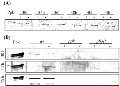
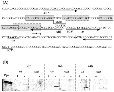
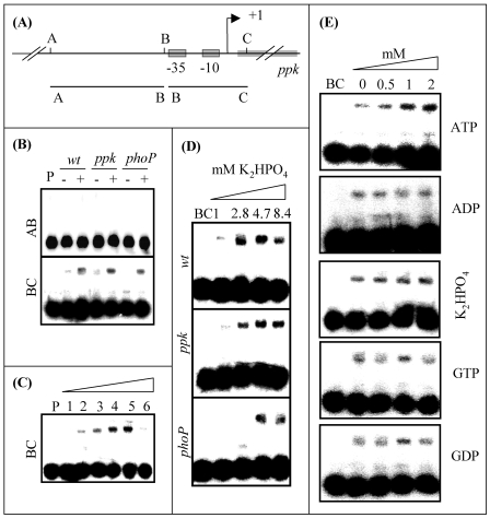
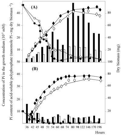
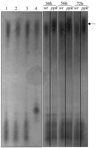
The ppk gene of Streptomyces lividans encodes an enzyme catalyzing, in vitro, the reversible polymerization of the gamma phosphate of ATP into polyphosphate and was previously shown to play a negative role in the control of antibiotic biosynthesis (H. Chouayekh and M. J. Virolle, Mol. Microbiol. 43:919-930, 2002). In the present work, some regulatory features of the expression of ppk were established and the polyphosphate content of S. lividans TK24 and the ppk mutant was determined. In Pi sufficiency, the expression of ppk was shown to be low but detectable. DNA gel shift experiments suggested that ppk expression might be controlled by a repressor using ATP as a corepressor. Under these conditions, short acid-soluble polyphosphates accumulated upon entry into the stationary phase in the wild-type strain but not in the ppk mutant strain. The expression of ppk under Pi-limiting conditions was shown to be much higher than that under Pi-sufficient conditions and was under positive control of the two-component system PhoR/PhoP. Under these conditions, the polyphosphate content of the cell was low and polyphosphates were reproducibly found to be longer and more abundant in the ppk mutant strain than in the wild-type strain, suggesting that Ppk might act as a nucleoside diphosphate kinase. In light of our results, a novel view of the role of this enzyme in the regulation of antibiotic biosynthesis in S. lividans TK24 is proposed.


Figures

FIG. 1.
FIG. 1.
(A) Western blot analysis of ppk expression in S. lividans TK24 grown on plates of modified minimal medium under phosphate-sufficient (+; 2.2 mM K2HPO4) or phosphate-limiting (−; 0.44 mM K2HPO4) conditions. Twenty-microgram portions of protein extract from mycelial samples taken after 30, 34, 36, 38, 40, and 44 h of growth were loaded into the wells. (B) Western blot analysis of ppk expression in S. lividans TK24 (wild type [wt]), S. lividans TK24 ppk::Ωhygro (ppk) and S. lividans TK24 phoP::Ωaac (phoP) grown under the conditions described above. Twenty-microgram portions of protein extracts from mycelial samples taken after 30, 44, and 66 h of growth were loaded into the wells. In panels A and B, 60 ng of purified His-tagged Ppk was loaded into the first well as control.
FIG. 2.
FIG. 2.
(A) Organization of the ppk promoter region. The ppk start codon is enclosed in a box. The −10 and −35 regions of ppk are underlined. The ppk transcriptional start site is indicated by an arrow. The region located upstream of the ppk promoter region was displaced from the promoter region by the insertion of an Ωaac cassette conferring resistance to apramycin at an EcoRI site created by PCR. The positions of the primers used for the amplification of the fragments, AB (131 bp) and BC (91 bp), used in band shift experiments shown in Fig. 3, are indicated by arrows. Some repeated motifs are enclosed in gray boxes. (B) Western blot analysis of ppk expression in S. lividans TK24 (wild type [wt]) and S. lividans TK24 EcoRI-Ωaacppk (mut) grown on modified minimal medium plates under Pi-sufficient (+; 2.2 mM K2HPO4) or Pi-limited (−; 0.44 mM K2HPO4) conditions. Twenty-microgram portions of protein extracts from mycelial samples taken after 30, 36, and 42 h of growth were loaded into the wells. Sixty nanograms of purified His-tagged Ppk was loaded into the first well as control.
FIG. 3.
FIG. 3.
(A) Schematic diagram of the ppk promoter region (B) Gel shift experiments carried out with DNA fragments of the ppk gene 5′ region. 32P-labeled PCR fragments AB and BC (0.5 ng/5,000 to 10,000 cpm) were incubated for 15 min at 30°C with 30-μg portions of protein extract of S. lividans TK24 (wild type [wt]), S. lividans TK24 ppk::Ωhygro (ppk), and S. lividans TK24 phoP::Ωaac (phoP) grown on solid R2YE medium under Pi-limited (−; 1 mM) or Pi-sufficient (+; 8.4 mM) conditions. (C) Gel-shift experiments carried out with fragment BC. 32P-labeled PCR fragment BC (0.5 ng/5,000 to 10,000 cpm) was incubated for 15 min at 30°C with 10, 20, 30, 40, or 50 μg of protein extracts (lanes 1 to 5, respectively) prepared from cultures of S. lividans TK24 grown for 44 h on solid R2YE medium containing 8.4 mM KH2PO4. For lane 6, 100 ng of the unlabeled fragment BC was added to 50 μg of protein extract. (D) Gel-shift experiments carried out with fragment BC in the presence of protein extracts prepared from cultures of S. lividans TK24 (wild type), S. lividans TK24 ppk::Ωhygro (ppk mutant) and S. lividans TK24 phoP::Ωaac (phoP mutant) grown for 44 h on solid R2YE medium containing 1, 2.85, 4.7, and 8.4 mM Pi, respectively. 32P-labeled PCR fragment BC (0.5 ng/5,000 to 10,000 cpm) was incubated for 15 min at 30°C with 30 μg of the various protein extracts. Free DNA and protein-bound DNA were separated in a 4% polyacrylamide gel as described in Materials and Methods. (E) Gel shift experiments carried out with 32P-labeled PCR fragment BC (0.5 ng/5,000 to 10,000 cpm) incubated for 15 min at 30°C with 30 μg of protein extract of S. lividans TK24 (wild type) grown on solid R2YE medium proficient in Pi (+; 4.7 mM) in the presence of various concentrations of ATP, ADP, GTP, GDP, or K2HPO4.
FIG. 4.
FIG. 4.
Growth curves for S. lividans TK24 (black diamonds) and S. lividans TK24 ppk::Ωhygro (white diamonds), phosphate contents of extracted acid-soluble polyphosphates from S. lividans TK24 (solid bars) and S. lividans TK24 ppk::Ωhygro (open bars), and concentrations of KH2PO4 in the growth medium throughout growth of S. lividans TK24 (black circles) and S. lividans TK24 ppk::Ωhygro (white circles) (A) Pi limited (R2YE medium containing 1 mM Pi). (B) Pi sufficient (R2YE medium containing 4.7 mM Pi).
FIG. 5.
FIG. 5.
Polyphosphates (indicated by an arrow) were purified on a silica gel as described in Materials and Methods from lawns of S. lividans TK24 (wild type [wt]) and S. lividans TK24 ppk::Ωhygro (ppk) grown for 36, 56, and 72 h on solid R2YE medium under Pi-limiting conditions (1 mM Pi). Polyphosphates purified from 36-h lawns of S. lividans TK24 either were not treated (lane 1) or were incubated for 4 h at 37°C with 0.25 μg/μl pronase (lane 2), 3 U/μl of RNase-free DNase I (lane 3), 1.25 μg/μl of RNase A (lane 3), and 2.5 U/μl of CIPA (lane 4). All polyphosphate samples were electrophoresed overnight on a 60-cm-long denaturating polyacrylamide gel stained with toluidine blue.
FIG. 6.
FIG. 6.
Growth curve and blue-pigmented antibiotic production (OD640) for S. lividans TK24 (black lozenges and solid bars) and S. lividans TK24 ppk::Ωhygro (white diamonds and open bars) grown on R2YE medium containing 1 mM phosphate (Pi-limiting conditions). A similar assay was done with the same strains grown on R2YE medium containing 4.7 mM phosphate (Pi-sufficient conditions), and no production of the blue-pigmented antibiotics was detected.

Similar articles

Cited by

References

    1. Ault-Riché, D., C. D. Fraley, C.-M. Tzeng, and A. Kornberg. 1998. Novel assay reveals multiple pathways regulating stress-induced accumulations of inorganic polyphosphate in Escherichia coli. J. Bacteriol. 180:1841-1847. - PMC - PubMed
    1. Ausubel, F. M., R. Brent, R. E. Kingston, D. D. Moore, J. G. Seidman, J. A. Smith, and K. Struhl. 1995. Current protocols in molecular biology. John Wiley & Sons, New York, N.Y.
    1. Avignone Rossa, C., J. White, A. Kuiper, P. W. Postma, M. Bibb, and M. J. Teixeira de Mattos. 2002. Carbon flux distribution in antibiotic-producing chemostat cultures of Streptomyces lividans. Metab. Eng. 4:138-150. - PubMed
    1. Bibb, M. J. 2005. Regulation of secondary metabolism in streptomycetes. Curr. Opin. Microbiol. 8:208-215. - PubMed
    1. Blondelet-Rouault, M. H., J. Weiser, A. Lebrihi, P. Branny, and J. L. Pernodet. 1997. Antibiotic resistance gene cassettes derived from the omega interposon for use in E. coli and Streptomyces. Gene 190:315-317. - PubMed

Publication types

MeSH terms