. 2006 Sep;188(17):6396-405.
doi: 10.1128/JB.00249-06.

Bacillus subtilis aconitase is required for efficient late-sporulation gene expression

Affiliations

Bacillus subtilis aconitase is required for efficient late-sporulation gene expression

Alisa W Serio et al. J Bacteriol. 2006 Sep.

Erratum in

  • J Bacteriol. 2007 Jul;189(14):5403

Abstract

Bacillus subtilis aconitase, encoded by the citB gene, is homologous to the bifunctional eukaryotic protein IRP-1 (iron regulatory protein 1). Like IRP-1, B. subtilis aconitase is both an enzyme and an RNA binding protein. In an attempt to separate the two activities of aconitase, the C-terminal region of the B. subtilis citB gene product was mutagenized. The resulting strain had high catalytic activity but was defective in sporulation. The defect was at a late stage of sporulation, specifically affecting expression of sigmaK-dependent genes, many of which are important for spore coat assembly and require transcriptional activation by GerE. Accumulation of gerE mRNA and GerE protein was delayed in the aconitase mutant strain. Pure B. subtilis aconitase bound to the 3' untranslated region of gerE mRNA in in vitro gel mobility shift assays, strongly suggesting that aconitase RNA binding activity may stabilize gerE mRNA in order to allow efficient GerE synthesis and proper timing of spore coat assembly.


Figures

FIG. 1.
FIG. 1.
C-terminal alignment of human IRP-1 and B. subtilis aconitase. Human IRP-1 residues in bold have been defined as important for IRP-1-RNA interaction (23). B. subtilis citB residues in bold are the correlating residues and were targeted for site-directed mutagenesis.
FIG. 2.
FIG. 2.
Specific activity of aconitase in cell extracts. AWS144 (citB+; closed circles) and AWS133 (citB5; closed triangles) were grown in DSM, and cell extracts were prepared at the time points indicated and analyzed for aconitase enzyme activity (open symbols).
FIG. 3.
FIG. 3.
RT-PCR analysis of gerE transcript levels. AWS144 (citB+; triangles) and AWS133 (citB5; circles) were grown in DSM at 37°C (inset). Samples were isolated at the time points indicated. RNA was prepared, and cDNA was synthesized with reverse primers to rrnA-16S and gerE. Eighteen cycles of amplification was determined to be within the linear range for PCR of rrnA-16S. The linear range for PCR of gerE was determined for each sample; 48 to 50 cycles were used for time points 6.5 to 8.5, and 35 cycles were used for time points 9 to 10.5. The rrnA-16S PCRs were done in duplicate for each sample. After product quantitation, the pixel numbers were averaged and normalized by determining the ratio of each sample to the wild-type rrnA-16S (h 6.5 sample). Then, all gerE transcripts were normalized to the rrnA-16S product from the same time point by determining the ratio of gerE product to the normalized rrnA-16S product. Relative gerE transcripts for AWS144 (citB+; solid bars) and AWS133 (citB5; empty bars) are shown for each time point.
FIG. 4.
FIG. 4.
Accumulation of GerE protein in mutant and wild-type (wt) strains. Extracts of stationary-phase cultures of strains AWS144 (citB+), AWS133 (citB5), and EUDC9901 (gerE::kan) harvested at the indicated time points (Tn = n h after the end of the exponential growth phase) were assayed for GerE protein by immunoblotting after separation by SDS-15% PAGE. GerE (8 kDa; indicated by the arrow) was the fastest-migrating band that reacted with antibody.
FIG. 5.
FIG. 5.
Expression of lacZ fusions to sporulation-specific promoters. Strains AWS154 (citB+ spoIIA-lacZ; circles) and AWS153 (citB5 spoIIA-lacZ; triangles) (top) or strains AWS145 (citB+ cotA-lacZ; circles) and AWS137 (citB5 cotA-lacZ; triangles) (bottom) were grown in DSM at 37°C. Samples were removed for measurement of the OD600 (closed symbols) and for assay of β-galactosidase activity (open symbols).
FIG. 6.
FIG. 6.
Immunoblot with antibody to σK. AWS144 (citB+ [wt]), AWS133 (citB5), and JHBS5 (citB+ ΔsigK [ΔK]) were grown in DSM at 37°C. Samples were isolated at the time points indicated (T, indicated number of hours after entry into stationary phase). Crude cellular extracts were subjected to SDS-PAGE and analyzed by immunoblotting with antibody specific to the σK protein.
FIG. 7.
FIG. 7.
Eukaryotic IRE consensus sequence and a putative stem-loop structure of the gerE mRNA 3′ UTR. The gerE putative stem-loop structure is found 54 bases after the translational stop codon.
FIG. 8.
FIG. 8.
Gel mobility shift assays of aconitase binding to gerE mRNA. The gerE mRNA 3′ UTR (A), the gerE antisense RNA 3′ UTR (B), and the fliT antisense RNA 3′ UTR (C) were synthesized and radiolabeled by in vitro transcription. Radiolabeled RNA was incubated without or with increasing concentrations of pure B. subtilis His10-aconitase, as indicated, and then analyzed for complex formation by gel mobility shift assay.
FIG. 9.
FIG. 9.
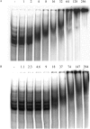
Gel mobility shift assays with partially purified aconitase. The gerE mRNA 3′ UTR was synthesized and radiolabeled by in vitro transcription. Radiolabeled RNA was incubated without or with increasing concentrations of wild-type aconitase-His6 (A) or citB5 mutant aconitase-His6 (B), as indicated, and then analyzed for complex formation by gel mobility shift assay. The amount of partially purified aconitase (aconitase units) added to each reaction was independently estimated by quantitative immunoblot analysis.

Similar articles

Cited by

References

    1. Addess, K. J., J. P. Basilion, R. D. Klausner, T. A. Rouault, and A. Pardi. 1997. Structure and dynamics of the iron responsive element RNA: implications for binding of the RNA by iron regulatory binding proteins. J. Mol. Biol. 274:72-83. - PubMed
    1. Alén, C., and A. L. Sonenshein. 1999. Bacillus subtilis aconitase is an RNA-binding protein. Proc. Natl. Acad. Sci. USA 96:10412-10417. - PMC - PubMed
    1. Basilion, J. P., T. A. Rouault, C. M. Massinople, R. D. Klausner, and W. H. Burgess. 1994. The iron-responsive element-binding protein: localization of the RNA-binding site to the aconitase active-site cleft. Proc. Natl. Acad. Sci. USA 91:574-578. - PMC - PubMed
    1. Bulteau, A. L., H. A. O'Neill, M. C. Kennedy, M. Ikeda-Saito, G. Isaya, and L. I. Szweda. 2004. Frataxin acts as an iron chaperone protein to modulate mitochondrial aconitase activity. Science 305:242-245. - PubMed
    1. Constable, A., S. Quick, N. K. Gray, and M. W. Hentze. 1992. Modulation of the RNA-binding activity of a regulatory protein by iron in vitro: switching between enzymatic and genetic function? Proc. Natl. Acad. Sci. USA 89:4554-4558. - PMC - PubMed

Publication types

MeSH terms

LinkOut - more resources