. 2006 Oct;26(20):7430-6.
doi: 10.1128/MCB.00624-06. Epub 2006 Aug 21.

Redox regulation of human OGG1 activity in response to cellular oxidative stress

Affiliations

Redox regulation of human OGG1 activity in response to cellular oxidative stress

Anne Bravard et al. Mol Cell Biol. 2006 Oct.

Abstract

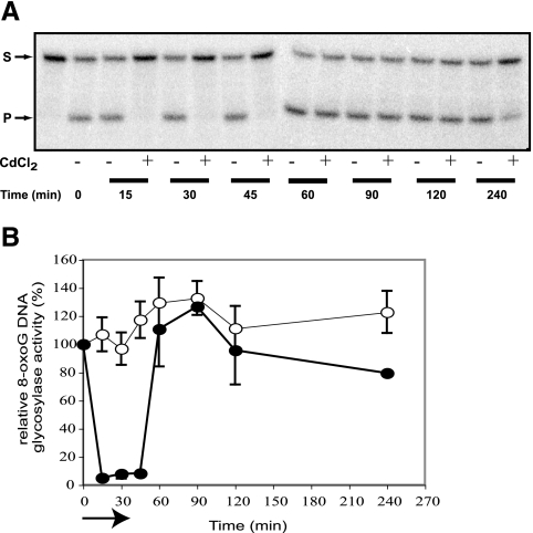
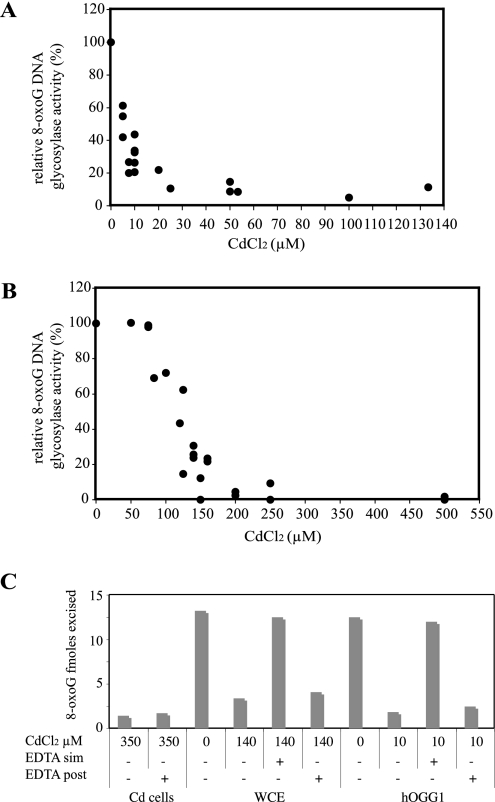
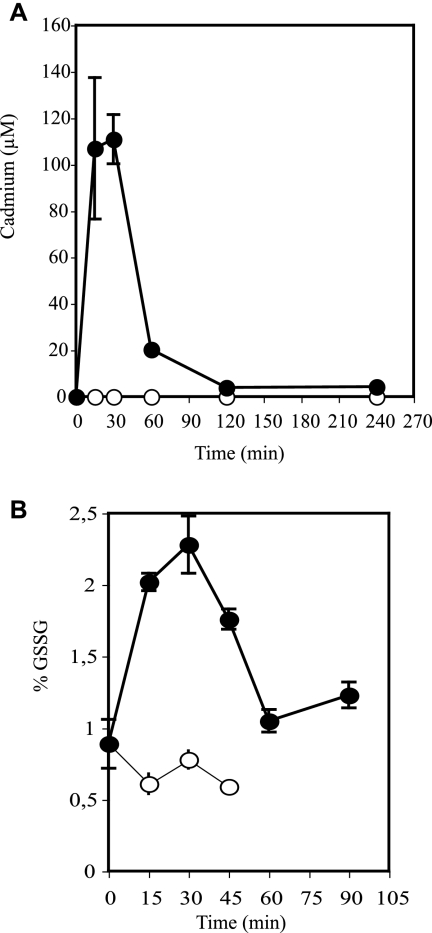
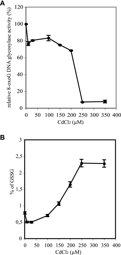
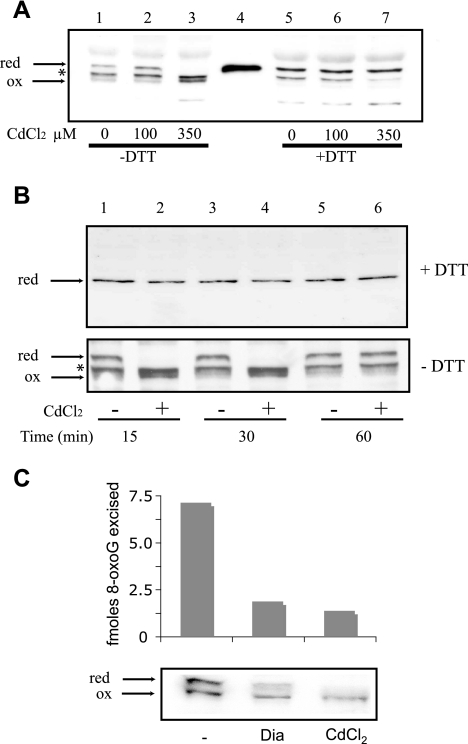
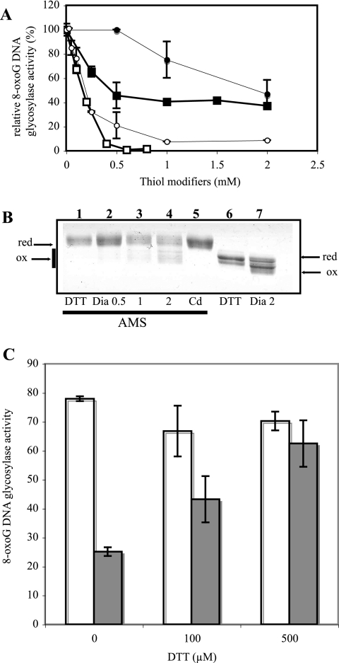
8-Oxoguanine (8-oxoG), a common and mutagenic form of oxidized guanine in DNA, is eliminated mainly through base excision repair. In human cells its repair is initiated by human OGG1 (hOGG1), an 8-oxoG DNA glycosylase. We investigated the effects of an acute cadmium exposure of human lymphoblastoid cells on the activity of hOGG1. We show that coinciding with alteration of the redox cellular status, the 8-oxoG DNA glycosylase activity of hOGG1 was nearly completely inhibited. However, the hOGG1 activity returned to normal levels once the redox cellular status was normalized. In vitro, the activity of purified hOGG1 was abolished by cadmium and could not be recovered by EDTA. In cells, however, the reversible inactivation of OGG1 activity by cadmium was strictly associated with reversible oxidation of the protein. Moreover, the 8-oxoG DNA glycosylase activity of purified OGG1 and that from crude extracts were modulated by cysteine-modifying agents. Oxidation of OGG1 by the thiol oxidant diamide led to inhibition of the activity and a protein migration pattern similar to that seen in cadmium-treated cells. These results suggest that cadmium inhibits hOGG1 activity mainly by indirect oxidation of critical cysteine residues and that excretion of the metal from the cells leads to normalization of the redox cell status and restoration of an active hOGG1. The results presented here unveil a novel redox-dependent mechanism for the regulation of OGG1 activity.


Figures

FIG. 1.
FIG. 1.
Reversible inhibition of the 8-oxoG DNA glycosylase activity in cadmium-treated cells. After a 30-min exposure to 350 μM CdCl2, cells were washed and returned to growth medium at 37°C. Cells were harvested at different times during and after treatment, and the 8-oxoG DNA glycosylase activity was determined on 10 μg of cell extracts as described in Materials and Methods. (A) Representative gel showing the cleavage of the 5′-end-labeled 34-mer duplex containing an 8-oxoG (S). The 16-mer product (P) is indicated by the second arrow. (B) Quantification of hOGG1 activities. Filled and open symbols correspond to extracts from cadmium-treated cells and corresponding control cells, respectively. The arrow indicates the treatment time. Data correspond to at least four experiments.
FIG. 2.
FIG. 2.
Direct and irreversible effect of cadmium on hOGG1 activity. (A and B) Purified hOGG1 (0.5 ng) (A) or extracts from Boleth control cells (10 μg) (B) were incubated for 15 min at 37°C with various amounts of cadmium chloride and then added to the glycosylase reaction mixture as described previously. (C) Effect of 5 mM EDTA on 8-oxoG DNA glycosylase activity in extracts (10 μg) from in vivo cadmium-treated cells (Cd cells), on extracts from in vitro cadmium-treated normal cell extracts (WCE), or on 0.5 ng of purified hOGG1 (hOGG1). In the cases of WCE or hOGG1 EDTA was added either simultaneously with cadmium (sim) or for 15 min after cadmium treatment (post) of the samples.
FIG. 3.
FIG. 3.
Kinetics of metal accumulation and of alteration of the glutathione balance in cadmium-exposed cells. Cells treated and collected as described for Fig. 1 were analyzed for intracellular cadmium concentrations by ICP-MS (A) and level of oxidized glutathione expressed as a percentage of the total glutathione content (B). Filled and open symbols correspond to extracts from cadmium-treated and control cells, respectively. Each point represents the average of at least four independent experiments ± standard deviation. Total glutathione content varied less than 20% between control and Cd-treated cells.
FIG. 4.
FIG. 4.
Dose effect of cadmium on cellular 8-oxoG DNA glycosylase activity and glutathione balance. Cells were harvested after 30 min of incubation at 37°C with various CdCl2 concentrations and analyzed for 8-oxoG DNA glycosylase activity as described for Fig. 1 (A) and content in oxidized glutathione (B). Data points represent the average of three independent determinations and their standard deviations.
FIG. 5.
FIG. 5.
Cadmium-induced reversible oxidation of the cellular hOGG1 protein. (A) Comparison between nonreductive (without DTT) and reductive (with 100 mM DTT) hOGG1 Western blot assays for protein extracts from cells exposed for 30 min at 100 μM and 350 μM compared to control cells. Lane 4 corresponds to 10 ng of a fully reduced form of hOGG1 obtained by denaturation of the protein in Laemmli buffer with 100 mM DTT. The middle band (*) either is a nonspecific band or reflects a nonredox modification of hOGG1 present in the extracts. (B) (Upper panel) Western blot assays for hOGG1 performed under standard (reducing) conditions on 50 μg of protein extracts from cells exposed for 30 min to 350 μM cadmium chloride and corresponding control cells, obtained at 15 min and 30 min during the metal treatment and 30 min thereafter. (Lower panel) The same extracts were analyzed for the presence of hOGG1 by Western blotting under nonreducing conditions, showing the reduced (upper band) and oxidized (lower band) forms of the protein. (C) (Upper panel) 8-OxoG DNA glycosylase activity on 12 μg of protein extracts from control cells (−) or cells treated for 30 min with either 2 mM diamide (Dia) or 350 μM CdCl2. (Lower panel) Western blot assay for hOGG1 under nonreducing conditions on 50 μg of protein from the same extracts.
FIG. 6.
FIG. 6.
Effects of thiol-modifying agents on hOGG1 activity and migration pattern. (A) Protein extracts (10 μg) from normal cells (squares) or purified hOGG1 (0.5 ng) (circles) were incubated for 15 min in 20 mM Tris-HCl, pH 6.8, containing various concentrations of NEM (open symbols) or diamide (filled symbols) and then analyzed for their 8-oxoG DNA glycosylase activity. Results are expressed as percentages of the control untreated samples. (B) Migration pattern of hOGG1 (750 ng) after 15 min of incubation with the following reagents: DTT (100 mM), diamide (Dia; 0.5, 1, or 2 mM), and Cd (0.5 mM). On the left of the gel (lanes 1 to 5) samples were incubated after treatment with 10 mM AMS to label free sulfhydryls. Lanes 6 and 7 show reactions under nonreducing conditions. The gel was color stained. (C) Effect of 100 and 500 μM DTT on the 8-oxoG DNA glycosylase activity of purified hOGG1 (5 ng) previously oxidized by 15 min of incubation in the presence of 2 mM diamide in 20 mM Tris-HCl, pH 6.8. The excess diamide was removed before DTT was added through two passages on 10K Microcon columns (Amicon). Open bars correspond to control experiments where the protein was not exposed to diamide.

References

    1. Anderson, M. E. 1985. Tissue glutathione, p. 317-323. In R. A. Greenwald (ed.), Handbook of methods for oxygen radical research. CRC Press, Inc., Boca Raton, Fla.
    1. Audebert, M., J. B. Charbonnier, S. Boiteux, and J. P. Radicella. 2002. Mitochondrial targeting of human 8-oxoguanine DNA glycosylase hOGG1 is impaired by a somatic mutation found in kidney cancer. DNA Repair (Amsterdam) 1:497-505. - PubMed
    1. Biswas, S., A. S. Chida, and I. Rahman. 2006. Redox modifications of protein-thiols: emerging roles in cell signaling. Biochem. Pharmacol. 71:551-564. - PubMed
    1. Bjoras, M., L. Luna, B. Johnsen, E. Hoff, T. Haug, T. Rognes, and E. Seeberg. 1997. Opposite base-dependent reactions of a human base excision repair enzyme on DNA containing 7,8-dihydro-8-oxoguanine and abasic sites. EMBO J. 16:6314-6322. - PMC - PubMed
    1. Boiteux, S., and J. P. Radicella. 2000. The human OGG1 gene: structure, functions, and its implication in the process of carcinogenesis. Arch. Biochem. Biophys. 377:1-8. - PubMed

Publication types