. 2006 Aug 29;103(35):13180-5.
doi: 10.1073/pnas.0605669103. Epub 2006 Aug 21.

Alveolar epithelial cell mesenchymal transition develops in vivo during pulmonary fibrosis and is regulated by the extracellular matrix

Affiliations

Alveolar epithelial cell mesenchymal transition develops in vivo during pulmonary fibrosis and is regulated by the extracellular matrix

Kevin K Kim et al. Proc Natl Acad Sci U S A. .

Abstract

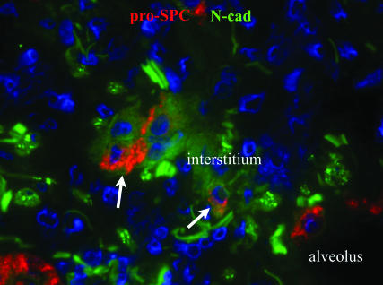
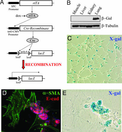
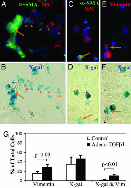
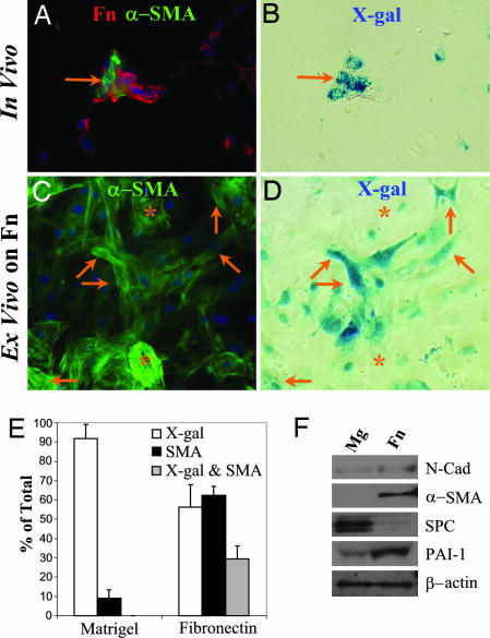
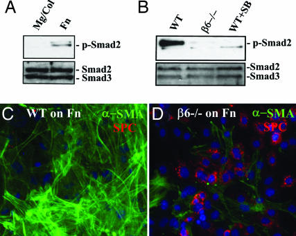
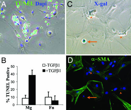
Mechanisms leading to fibroblast accumulation during pulmonary fibrogenesis remain unclear. Although there is in vitro evidence of lung alveolar epithelial-to-mesenchymal transition (EMT), whether EMT occurs within the lung is currently unknown. Biopsies from fibrotic human lungs demonstrate epithelial cells with mesenchymal features, suggesting EMT. To more definitively test the capacity of alveolar epithelial cells for EMT, mice expressing beta-galactosidase (beta-gal) exclusively in lung epithelial cells were generated, and their fates were followed in an established model of pulmonary fibrosis, overexpression of active TGF-beta1. beta-gal-positive cells expressing mesenchymal markers accumulated within 3 weeks of in vivo TGF-beta1 expression. The increase in vimentin-positive cells within injured lungs was nearly all beta-gal-positive, indicating epithelial cells as the main source of mesenchymal expansion in this model. Ex vivo, primary alveolar epithelial cells cultured on provisional matrix components, fibronectin or fibrin, undergo robust EMT via integrin-dependent activation of endogenous latent TGF-beta1. In contrast, primary cells cultured on laminin/collagen mixtures do not activate the TGF-beta1 pathway and, if exposed to active TGF-beta1, undergo apoptosis rather than EMT. These data reveal alveolar epithelial cells as progenitors for fibroblasts in vivo and implicate the provisional extracellular matrix as a key regulator of epithelial transdifferentiation during fibrogenesis.


Conflict of interest statement

Conflict of interest statement: No conflicts declared.

Figures

Fig. 1.
Fig. 1.
Evidence of early EMT in IPF lung. IPF lung biopsy demonstrating cells removed from the alveolar basement membrane within the interstitium coexpressing pro-SPC and the mesenchymal protein, N-cadherin (arrows).
Fig. 2.
Fig. 2.
Characterization of reporter mice expressing AEC β-gal. (A) Triple transgenic mice express rtTA in lung epithelial cells using the SPC promoter. In the presence of doxycycline (dox), rtTA is an active transcriptional factor leading to expression of Cre recombinase and removal of the floxed poly(A) sequence in the ROSA allele. Thus, in cells expressing SPC, expression of lacZ is constitutively and irreversibly activated. (B) Immunoblot of lysate from different tissues demonstrating lung-specific expression of β-gal. (C) Expression of β-gal in a lung section by X-gal staining. (D and E) Type II AECs isolated from triple transgenic mice, maintained on Mg for 1 week, then stained with X-gal (E) followed by immunostaining for E-cadherin and α-SMA (D). X-gal-positive epithelial cells form clusters and stain for E-cadherin. Contaminating fibroblasts are X-gal-negative and α-SMA-positive.
Fig. 3.
Fig. 3.
EMT develops in vivo after TGF-β1-mediated lung injury. (A and B) The same field (×60) of a lung 3 weeks after intranasal AdTGF-β1 stained by X-gal (B) then immunostained (A) for α-SMA and pro-SPC. Several X-gal-positive, pro-SPC-positive cells are demonstrated (arrowheads) as well as a cluster of X-gal-positive, α-SMA-positive, and pro-SPC-negative cells (arrow). (C and D) The same field of a whole lung single-cell suspension from an injured lung stained by X-gal (D) then immunostained (C) for α-SMA and pro-SPC. An X-gal-positive, α-SMA-positive, pro-SPC-negative cell is demonstrated (arrow). (E and F) The same field of a whole lung single cell suspension from a triple transgenic mouse three weeks after AdTGF-β1, stained by X-gal (F), and immunostained (E) for vimentin. An X-gal- and vimentin-positive cell is indicated by arrow. (G) Quantification X-gal- and vimentin-positive single cells from of AdTGF-β1-injured and control lungs (n = 3 per group).
Fig. 4.
Fig. 4.
Fn drives AEC EMT ex vivo. (A and B) The same field (×60) of a lung 3 weeks after intranasal AdTGF-β1 stained by X-gal (B), then immunostained (A) for α-SMA (green) and Fn (red). A cluster of X-gal- and α-SMA-positive cells within an area of Fn deposition is demonstrated (arrow). (C and D) The same view of isolated AECs from an uninjured triple transgenic mouse cultured on Fn-coated slides for 1 week, then stained with X-gal (D) then immunostained (C) for α-SMA. Several X-gal- and α-SMA-positive cells are shown (arrows) as well as several X-gal-negative, α-SMA-positive cells (∗). (E) Quantification of primary AECs from triple transgenic mice maintained on Fn- or Mg-coated slides for 1 week and stained for X-gal and α-SMA. (F) Primary murine AECs were cultured on Fn- or Mg-coated plates for 2 days and then lysed and analyzed by immunoblot.
Fig. 5.
Fig. 5.
AEC EMT requires αvβ6-dependent latent TGF-β1 activation. (A) Primary murine AECs were cultured for 2 days on Mg/Col or Fn, then lysed and analyzed by immunoblot for p-Smad2 and total Smad2/3. (B) Primary AECs from wild-type and α6-null mice were cultured on Fn ± SB431542 (10 μM) for 2 days, then lysed and immunoblotted for p-Smad2 and total Smad2/3. (C and D) AECs from wild-type (C) and β6-null (D) mice were cultured on Fn for 4 days, then stained for α-SMA and pro-SPC.
Fig. 6.
Fig. 6.
TGF-β1-induced AEC apoptosis on Mg but not Fn. (A) Overlay of phase with TUNEL (green) and Dapi (blue) staining of AECs plated on Mg and treated with TGF-β1 (4 ng/ml) for 2 days. (B) Quantification of TUNEL-positive cells. (C and D) AECs from triple transgenic mice cultured on Mg then stimulated with TGF-β1 for 1 week, stained with X-gal (C) followed by immunostaining for α-SMA (D). A remaining X-gal-positive cell is shown (arrow) as well as several apoptotic remnants (arrow heads).

References

    1. American Thoratic Society & European Respiratory Society. Am. J. Respir. Crit. Care Med. 2002;165:277–304. - PubMed
    1. Kalluri R., Neilson E. G. J. Clin. Invest. 2003;112:1776–1784. - PMC - PubMed
    1. Phillips R. J., Burdick M. D., Hong K., Lutz M. A., Murray L. A., Xue Y. Y., Belperio J. A., Keane M. P., Strieter R. M. J. Clin. Invest. 2004;114:438–446. - PMC - PubMed
    1. Moore B. B., Kolodsick J. E., Thannickal V. J., Cooke K., Moore T. A., Hogaboam C., Wilke C. A., Toews G. B. Am. J. Pathol. 2005;166:675–684. - PMC - PubMed
    1. Zeisberg M., Hanai J., Sugimoto H., Mammoto T., Charytan D., Strutz F., Kalluri R. Nat. Med. 2003;9:964–968. - PubMed

Publication types

MeSH terms