. 2006 Aug 29;103(35):13045-50.
doi: 10.1073/pnas.0601149103. Epub 2006 Aug 21.

Surface-modulated motion switch: capture and release of iron-sulfur protein in the cytochrome bc1 complex

Affiliations

Surface-modulated motion switch: capture and release of iron-sulfur protein in the cytochrome bc1 complex

Lothar Esser et al. Proc Natl Acad Sci U S A. .

Abstract

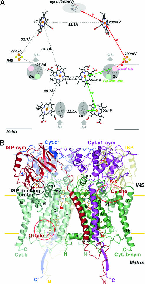
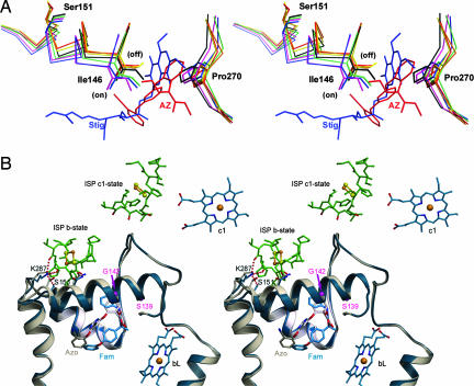
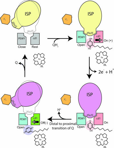
In the cytochrome bc(1) complex, the swivel motion of the iron-sulfur protein (ISP) between two redox sites constitutes a key component of the mechanism that achieves the separation of the two electrons in a substrate molecule at the quinol oxidation (Q(o)) site. The question remaining is how the motion of ISP is controlled so that only one electron enters the thermodynamically favorable chain via ISP. An analysis of eight structures of mitochondrial bc(1) with bound Q(o) site inhibitors revealed that the presence of inhibitors causes a bidirectional repositioning of the cd1 helix in the cytochrome b subunit. As the cd1 helix forms a major part of the ISP binding crater, any positional shift of this helix modulates the ability of cytochrome b to bind ISP. The analysis also suggests a mechanism for reversal of the ISP fixation when the shape complementarity is significantly reduced after a positional reorientation of the reaction product quinone. The importance of shape complementarity in this mechanism was confirmed by functional studies of bc(1) mutants and by a structure determination of the bacterial form of bc(1). A mechanism for the high fidelity of the bifurcated electron transfer is proposed.


Conflict of interest statement

Conflict of interest statement: No conflicts declared.

Figures

Fig. 1.
Fig. 1.
Prosthetic groups and subunit structures of the cyt bc1 complex. (A) Arrangement of prosthetic groups in the dimeric bc1 complex and illustration of the electron bifurcation at the Qo site. The bL, bH, and c1 heme groups are shown as ball-and-stick models, and the [2Fe2S] clusters are depicted as cpk models. Carbon atoms, black; nitrogen, blue; oxygen, red; sulfur, yellow; iron, brown. The Qo pockets near the IMS side of the membrane and the Qi pockets near the matrix side are labeled and shaded in gray. Cyt c is shown as a gray shaded oval. Distances between redox centers are given on the left half of the diagram, and the redox potential for each center is given on the right. The high- and low-potential ET paths are depicted with red and green arrows, respectively. Circles in pink and light green within the Qo pockets are hypothesized distal-QH2 and proximal-Q binding sites, respectively. (B) Ribbon diagram of the dimeric cyt b, cyt c1, and ISP subunit in the mitochondrial bc1 complex. Two symmetry-related cyt b subunits are shown (green and light green). The eight TM helices of cyt b are denoted with letters A–H. Helices A–E form one bundle in which the two b-type hemes (bL and bH in ball-and-stick models) reside; helices F–H form the other bundle. The ISP subunit (yellow and red for the symmetry pair) has an extrinsic soluble domain with a [2Fe2S] cluster at its tip, connecting to a TM segment by a flexible neck. The extrinsic domain of cyt c1 (blue and magenta for the symmetry pair) with its heme group is rigidly attached to its TM helix. The locations for the two active sites (Qo and Qi) per monomer in cyt b are labeled. The surface depression in cyt b at the IMS side of the membrane is labeled as the ISP-docking crater.
Fig. 2.
Fig. 2.
Motions observed in the cd1 helix and in the ef loop in the presence of two types of inhibitors. (A) Impact of inhibitors on the conformation of the cd1 switch at the Qo site are illustrated by this stereoscopic pair. Part of the cd1 helix and the P270 of the PEWY motif with and without bound inhibitors are shown by the stick models. Colors are as follows: native (black), stigmatellin (blue), famoxadone (green), UHDBT (magenta), azoxystrobin (red), myxothiazol (orange), and MOAS (yellow). Upon binding of different types of inhibitors, such as stigmatellin (Stig; blue molecule) or azoxystrobin (AZ; red molecule), I146 along with the entire cd1 helix moves either into the on or off position. (B) Stereo pair showing the structural features of the cd1 switch in ribbon form. The structure with famoxadone (Fam) is in blue, and that with azoxystrobin (Azo) is in gray. The inhibitors azoxystrobin and famoxadone are shown as stick models and as labeled. The side chains of K287 and S151 are shown in stick models with the dashed lines indicating H-bonds. H-bonds are also formed between K287 and residues from the ISP-ED when the cd1 switch is at the on position (blue model). In the native structure, or in inhibitor structures in which the ISP-ED is in a free or c1-state (8), no H-bond is formed between K287 and S151 (gray model).
Fig. 3.
Fig. 3.
Control of the ISP-ED motion switch and the proposed mechanism for electron bifurcation at the Qo pocket. The structural components necessary for the control of ISP conformational switch are illustrated in this cartoon rendition of the Qo pocket. The PEWY motif and cd1 helix in gray represent a native (Rest) configuration. The ISP in yellow and magenta are of oxidized and reduced form, respectively. The PEWY in blue stands for the open configuration with a bound Qo site inhibitor. The cd1 helix in red symbolizes the conformation (On/+) in the presence of a Pf inhibitor occupying the distal site (pink), and the cd1 helix in green shows the conformation (Off/−) when a Pm inhibitor is taking the proximal site (purple). Cyt c1 and heme bL are also shown.

References

    1. Trumpower B. L. J. Biol. Chem. 1990;265:11409–11412. - PubMed
    1. DiMauro S., Schon E. A. N. Engl. J. Med. 2003;348:2656–2668. - PubMed
    1. Staniek K., Gille L., Kozlov A. V., Nohl H. Free Radical Res. 2002;36:381–387. - PubMed
    1. Brasseur G., Saribas A. S., Daldal F. Biochim. Biophys. Acta. 1996;1275:61–69. - PubMed
    1. Bartlett D. W., Clough J. M., Godwin J. R., Hall A. A., Hamer M., Parr-Dobrzanski B. Pest Manag. Sci. 2002;58:649–662. - PubMed

Publication types

MeSH terms