. 2006 Oct 15;99(3):671-8.
doi: 10.1002/jcb.20982.

Downregulation of miR-122 in the rodent and human hepatocellular carcinomas

Affiliations

Downregulation of miR-122 in the rodent and human hepatocellular carcinomas

Huban Kutay et al. J Cell Biochem. .

Retraction in

Abstract

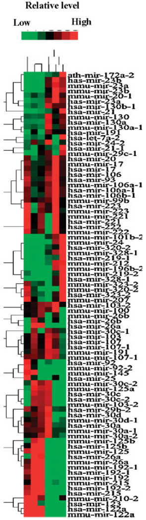
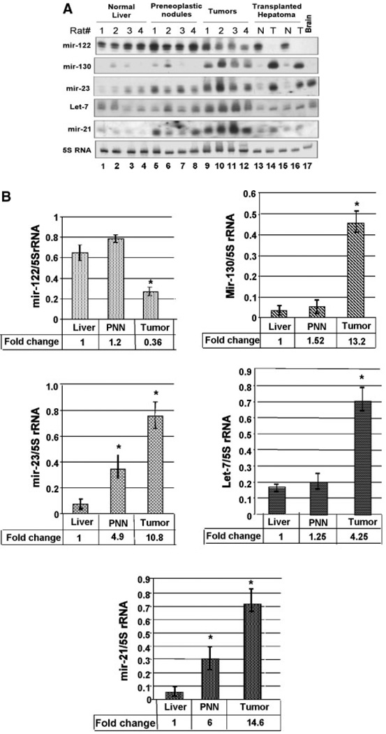
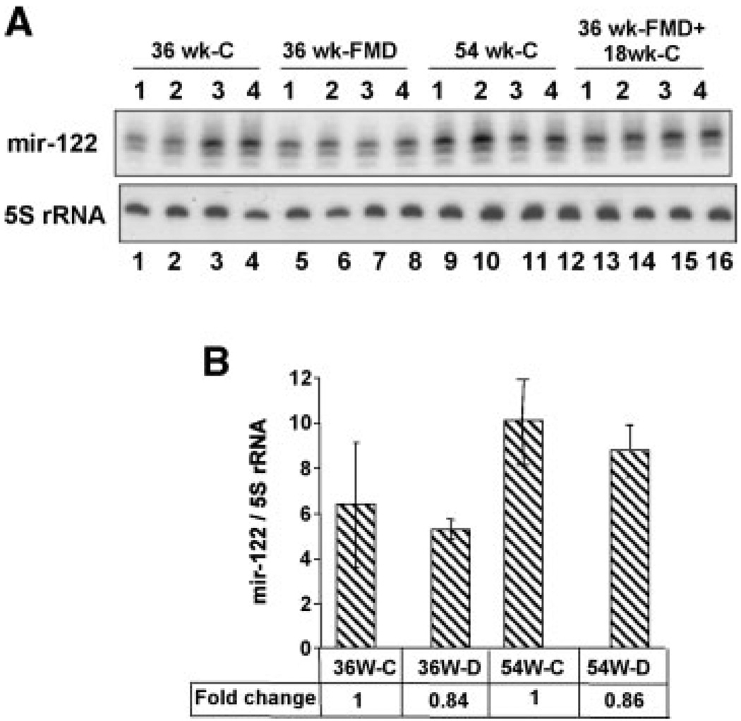
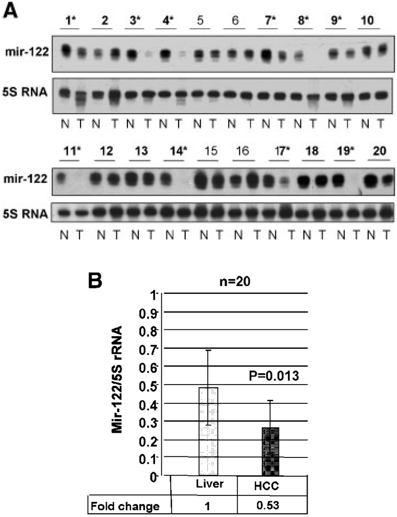
MicroRNAs (miRs) are conserved small non-coding RNAs that negatively regulate gene expression. The miR profiles are markedly altered in cancers and some of them have a causal role in tumorigenesis. Here, we report changes in miR expression profile in hepatocellular carcinomas (HCCs) developed in male Fisher rats-fed folic acid, methionine, and choline-deficient (FMD) diet. Comparison of the miR profile by microarray analysis showed altered expression of some miRs in hepatomas compared to the livers from age-matched rats on the normal diet. While let-7a, miR-21, miR-23, miR-130, miR-190, and miR-17-92 family of genes was upregulated, miR-122, an abundant liver-specific miR, was downregulated in the tumors. The decrease in hepatic miR-122 was a tumor-specific event because it did not occur in the rats switched to the folate and methyl-adequate diet after 36 weeks on deficient diet, which did not lead to hepatocarcinogenesis. miR-122 was also silent in a transplanted rat hepatoma. Extrapolation of this study to human primary HCCs revealed that miR-122 expression was significantly (P = 0.013) reduced in 10 out of 20 tumors compared to the pair-matched control tissues. These findings suggest that the downregulation of miR-122 is associated with hepatocarcinogenesis and could be a potential biomarker for liver cancers.


Figures

Fig. 1
Fig. 1
Cluster analysis of three HCCs and three age-matched rat livers. Microarray analysis of miRs expressed in the rat livers and hepatocellular carcinomas (HCCs). miR expression data were normalized to the average median of all the genes in the array. Only those miRs were used for clustering whose normalized value are higher than 1.5 and their expression were significantly different (ratio=1.5, OR=0.5) in at least one HCC and age-matched rat liver. N and T stands for normal liver and tumor, respectively.
Fig. 2
Fig. 2
A: Northern blot analysis confirmed downregulation of miR-122 and upregulation of miR-23, -21 -130, and let-7 in HCC compared with the controls. Total RNA was isolated from the livers of four rats-fed normal diet, four rats-fed FMD diet for 36 and 54 weeks, respectively. An aliquot (30 µg) of the total RNA was separated by denaturing PAGE, transferred to a nylon membrane and subjected to Northern blot analysis with 32P-labeled deoxyoligonucleotide antisense to specific miRs. The blot was reprobed with oligo antisense to 5S rRNA and the ratio of miR signals to that of 5S rRNA were determined. B: Quantitative analysis of Northern blot data showed significant downregulation of miR-122 and upregulation of miR-21, -23, and -130 and let-7 in rat HCCs. 32P-signal was measured using Imagequant software and quantified using volume analysis program. PNN stands for preneoplastic nodules. The results represent the average signal of each miR normalized to that of 5S rRNA ± SD. P-value of ≤0.05 was considered significant. Asterisks indicate changes that are significant.
Fig. 3
Fig. 3
A: Downregulation of miR-122 does not occur in the livers of rats switched to folate/methyl-adequate diet after 36 weeks on the FMD diet. The control animals were on adequate diet for 54 weeks whereas deficient animals were on FMD diet for the same time period. The third group was provided FMD diet for 36 weeks followed by adequate diet for another 18 weeks. Total RNA isolated from the livers was analyzed by Northern blotting. B: The quantitative representation of the data in (A). The results represent the average signal of each miR normalized to that of 5S rRNA ± SD. P-value of ≤0.05 was considered significant. The ratio of miR-122 to 5S rRNA in the controls (36 weeks-C and 54 weeks-C) was assigned a value of 1.
Fig. 4
Fig. 4
Reduced expression of miR-122 in human primary HCCs. A: An aliquot (5 µg) of total RNA from tumor (T) and matching normal (N) tissues was subjected to Northern blot analysis. Asterisks denote human primary HCCs in which miR-122 is downregulated. The signal of miR-122 normalized to that of 5S rRNA is presented below each sample. Asterisks indicate HCCs with significant decrease in miR-122. B: Quantitative analysis in all 20 samples showed that the decrease in miR-122 in HCCs was statistical significant.

References

    1. Ambros V. The functions of animal microRNAs. Nature. 2004;431:350–355. - PubMed
    1. Bartel DP. MicroRNAs: Genomics, biogenesis, mechanism, and function. Cell. 2004;116:281–297. - PubMed
    1. Calin GA, Sevignani C, Dumitru CD, Hyslop T, Noch E, Yendamuri S, Shimizu M, Rattan S, Bullrich F, Negrini M, Croce CM. Human microRNA genes are frequently located at fragile sites and genomic regions involved in cancers. Proc Natl Acad Sci USA. 2004;101:2999–3004. Epub 2004 Feb 18. - PMC - PubMed
    1. Chang J, Nicolas E, Marks D, Sander C, Lerro A, Bnendra MA, Xu C, Mason WS, Moloshok T, Bort R, Zaret KS, Taylor JM. Mir-122, a mammalian liver-specific microRNA, is processed from hcr mRNA and may down regulate the high affinity cationic amino acid transporter CAT-1. RNA Biology. 2004;1:106–113. - PubMed
    1. Du T, Zamore PD. microPrimer: The biogenesis and function of microRNA. Development. 2005;132:4645–4652. - PubMed

Publication types