Review
. 2006 Sep 15;398(3):319-37.
doi: 10.1042/BJ20060450.

Mechanisms of RecQ helicases in pathways of DNA metabolism and maintenance of genomic stability

Affiliations
Review

Mechanisms of RecQ helicases in pathways of DNA metabolism and maintenance of genomic stability

Sudha Sharma et al. Biochem J. .

Abstract

Helicases are molecular motor proteins that couple the hydrolysis of NTP to nucleic acid unwinding. The growing number of DNA helicases implicated in human disease suggests that their vital specialized roles in cellular pathways are important for the maintenance of genome stability. In particular, mutations in genes of the RecQ family of DNA helicases result in chromosomal instability diseases of premature aging and/or cancer predisposition. We will discuss the mechanisms of RecQ helicases in pathways of DNA metabolism. A review of RecQ helicases from bacteria to human reveals their importance in genomic stability by their participation with other proteins to resolve DNA replication and recombination intermediates. In the light of their known catalytic activities and protein interactions, proposed models for RecQ function will be summarized with an emphasis on how this distinct class of enzymes functions in chromosomal stability maintenance and prevention of human disease and cancer.


Figures

Figure 1
Figure 1. Schematic representation of RecQ helicase family members
Human members of the RecQ family of DNA helicases are shown. Proteins are aligned by their conserved RecQ helicase domain. The conserved domains and motifs in each group are shown by different colours as depicted at the bottom.
Figure 2
Figure 2. Conserved nuclease domain of WRN and 3′ to 5′ exonucleases
The top panel depicts the three conserved exonuclease motifs of human WRN and its homologues in lower eukaryotes. The two middle panels depict human DNA polymerases and exonucleases which share identity with WRN exonuclease. The bottom panel depicts a prokaryotic DNA polymerase with the conserved exonuclease domains. The conserved exonuclease domains are colour-coded as indicated at the bottom. Mm, Mus musculus.
Figure 3
Figure 3. Structures of conserved RecQ protein domains
(A) X-ray crystal structure of the E. coli RecQ catalytic core [12]. Helicase lobes are coloured red and blue, the Zn2+-binding domain and winged helix domain of the RQC region are in yellow and green respectively. A bound Zn2+ ion is shown as a magenta sphere. (B) Close-up of the ATP-binding site in the structure of the ATP[S]-bound E. coli RecQ catalytic core [7,12]. A bound Mn2+ ion is shown as a cyan sphere. (C) X-ray crystal structure of the E. coli RecQ HRDC domain [28]. Figures are courtesy of Dr James Keck (University of Wisconsin Medical School, Madison, WI, U.S.A.).
Figure 4
Figure 4. WRN exonuclease structure
(A) X-ray crystal structure of the WRN-exo fold is shown with the conserved active site residues that chelate the two Mn2+ ions (purple) [35]. (B) The WRN-exo hexameric ring model with uniquely coloured WRN-exo subunits was built by superimposition with the A. thaliana homologue [35]. Figures are courtesy of Dr John Tainer (The Scripps Research Institute, La Jolla, CA, U.S.A.).
Figure 5
Figure 5. Models depicting active unwinding mechanisms for DNA helicases
For all of the mechanisms depicted, DNA unwinding is fuelled by the energy of helicase-catalysed NTP hydrolysis. (A) Inchworm mechanism for a monomeric helicase in which the enzyme undergoes energetic and conformational changes to translocate unidirectionally along DNA and destabilize the duplex. (B) Co-operative inchworm mechanism characterized by the alignment of enzyme molecules to promote forward movement and increase helicase activity when multiple helicase monomers co-operate. (C) Rolling model mechanism in which the protomers of a dimeric helicase alternate in binding ssDNA or dsDNA as the enzyme translocates and destabilizes the duplex. (D) Hexameric helicase unwinding mechanism characterized by a ring structure that encircles one strand and displaces the other strand which is extruded outside the ring.
Figure 6
Figure 6. Potential functions of RecQ helicases at replication forks
Biochemical and cellular studies have suggested that RecQ helicases act upon certain DNA structural intermediates at the replication fork to ensure accurate processing and genome fidelity. RecQ helicases may act upon the replication fork in various capacities (resolution of secondary structures, lagging strand processing, branch fork migration, DNA-damage sensing or bypass, replication fork regression and WRN proofreading function). See the text for details.
Figure 7
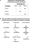
Figure 7. Interactions of RecQ helicases with structure-specific nucleases
(A) Reported physical and functional RecQ helicase interactions with human FEN-1, EXO-1 and Mus81 nucleases. ND, not determined. (B) Structural DNA intermediates of replication, recombination and repair that are acted upon by the concerted action of a RecQ helicase and a structure-specific nuclease. See the text for details.
Figure 8
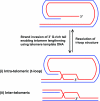
Figure 8. Proposed roles of RecQ helicases in telomere maintenance
T-loops are created through strand invasion of the 3′ telomeric overhang into the duplex region of the telomere. ALT is a recombination-based pathway that operates in telomerase-deficient cells. This pathway might involve RecQ- and TRF2-mediated strand invasion of the 3′ G-rich tail, enabling the telomere to be lengthened by using telomeric DNA as a template. The telomeric template DNA can originate from two sources: (i) intra-telomeric, where the t-loop is used to prime DNA synthesis, or (ii) inter-telomeric, where the DNA is copied from another telomere or telomeric sequence that exists as extrachromosomal DNA. Additionally, maintenance of telomere length might require a RecQ helicase (e.g. BLM or WRN) to resolve the D-loop structure at the t-loop or unwind a G-quadruplex in the 3′ G-rich tail.

References

    1. Harrigan J. A., Bohr V. A. Human diseases deficient in RecQ helicases. Biochimie. 2003;85:1185–1193. - PubMed
    1. Agrelo R., Cheng W. H., Setien F., Ropero S., Espada J., Fraga M. F., Herranz M., Paz M. F., Sanchez-Cespedes M., Artiga M. J., et al. Epigenetic inactivation of the premature aging Werner syndrome gene in human cancer. Proc. Natl. Acad. Sci. U.S.A. 2006;103:8822–8827. - PMC - PubMed
    1. Siitonen H. A., Kopra O., Kaariainen H., Haravuori H., Winter R. M., Säämänen A. M., Peltonen L., Kestila M. Molecular defect of RAPADILINO syndrome expands the phenotype spectrum of RECQL diseases. Hum. Mol. Genet. 2003;12:2837–2844. - PubMed
    1. Van Maldergem L., Siitonen H. A., Jalkh N., Chouery N., De Roy M., Delague V., Muenke M., Jabs E. W., Cai J., Wang L. L., et al. Revisiting the craniosynostosis-radial ray hypoplasia association: Baller–Gerold syndrome caused by mutations in the RECQL4 gene. J. Med. Genet. 2006;43:148–152. - PMC - PubMed
    1. Kyng K. J., May A., Kolvraa S., Bohr V. A. Gene expression profiling in Werner syndrome closely resembles that of normal aging. Proc. Natl. Acad. Sci. U.S.A. 2003;100:12259–12264. - PMC - PubMed

Publication types