. 2007 May;1773(5):642-52.
doi: 10.1016/j.bbamcr.2006.07.001. Epub 2006 Jul 14.

Regulation of the actin cytoskeleton in cancer cell migration and invasion

Affiliations

Regulation of the actin cytoskeleton in cancer cell migration and invasion

Hideki Yamaguchi et al. Biochim Biophys Acta. 2007 May.

Abstract

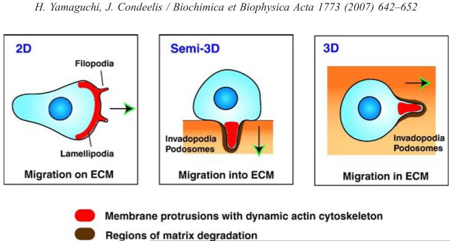
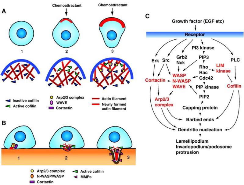
Malignant cancer cells utilize their intrinsic migratory ability to invade adjacent tissues and the vasculature, and ultimately to metastasize. Cell migration is the sum of multi-step processes initiated by the formation of membrane protrusions in response to migratory and chemotactic stimuli. The driving force for membrane protrusion is localized polymerization of submembrane actin filaments. Recently, several studies revealed that molecules that link migratory signals to the actin cytoskeleton are upregulated in invasive and metastatic cancer cells. In this review, we summarize recent progress on molecular mechanisms of formation of invasive protrusions used by tumor cells, such as lamellipodia and invadopodia, with regard to the functions of key regulatory proteins of the actin cytoskeleton; WASP family proteins, Arp2/3 complex, LIM-kinase, cofilin, and cortactin.


Figures

Fig. 1
Fig. 1
Cell migration and membrane protrusions in different environments. Cells migrating on 2D substrates form membrane protrusions called filopodia and lamellipodia at the leading edge. Cells entering into and migrating in a dense rigid ECM in 3D, such as tumor cells on top of a thick ECM and those found around blood vessels, need to form membrane protrusions at the invading front, such as invadopodia and podosomes that have an ECM remodeling activity. Formation of these structures is driven by localized actin polymerization. Proteins involved in formation of these protrusions are often upregulated in malignant cancer cells and associated with increased cell motility and invasion. Arrows indicate the direction of cell migration.
Fig. 2
Fig. 2
Model for lamellipodium and invadopodium/podosome formation. (A) 1: Unstimulated cells have non-polarized cell morphology in which molecular machinery for barbed end formation including cofilin is inactive. 2: Chemoattractant stimulation induces local activation of cofilin at the leading edge, which leads to severing of pre-existing actin filaments and formation of free barbed ends from which new actin filaments are assembled. This initiates membrane protrusions and sets the direction of cell migration. 3: Arp2/3 complex and WAVEs associate with newly formed actin filaments and induce formation of further barbed ends and the branched actin network. Subsequently, the branched actin filaments are stabilized by cortactin. This strengthens the protrusive force of lamellipodia and leads to cell movement. (B) 1: Invadopodium/podosome formation is triggered by N-WASP/WASP, Arp2/3 complex and cortactin, probably by coupled activation of growth factor receptor and integrin signaling. 2: This precursor is stabilized by further recruitment of invadopodium/podosome components and formation of actin network by cofilin. 3: Anchored precursor then gathers matrix-degrading proteinases to degrade ECM and protrude into matrix. N-WASP/Arp2/3 complex, cortactin, and cofilin continue to induce actin polymerization to maintain the structural core. In contrast to lamellipodia, the structure and organization of actin filaments are not yet determined in invadopodia/podosomes. (C) The signaling pathways leading to protrusion of lamellipodia and invadopodia/podosomes in response to growth factor stimulation. Molecules discussed in this review are highlighted in red.

References

    1. Condeelis J, Singer R, Segall JE. The great escape: when cancer cells hijack the genes for chemotaxis and motility. Annu. Rev. Cell Dev. Biol. 2005;21:695–718. - PubMed
    1. Sahai E. Mechanisms of cancer cell invasion. Curr. Opin. Genet. Dev. 2005;15:87–96. - PubMed
    1. Yamaguchi H, Wyckoff J, Condeelis J. Cell migration in tumors. Curr. Opin. Cell Biol. 2005;17:559–564. - PubMed
    1. Bailly M, Condeelis J. Cell motility: insights from the backstage. Nat. Cell Biol. 2002;4:E292–E294. - PubMed
    1. Pollard TD, Borisy GG. Cellular motility driven by assembly and disassembly of actin filaments. Cell. 2003;112:453–465. - PubMed

Publication types

LinkOut - more resources