. 2006 Sep;74(9):5067-74.
doi: 10.1128/IAI.00182-06.

Infection of human endothelial cells with spotted Fever group rickettsiae stimulates cyclooxygenase 2 expression and release of vasoactive prostaglandins

Affiliations

Infection of human endothelial cells with spotted Fever group rickettsiae stimulates cyclooxygenase 2 expression and release of vasoactive prostaglandins

Elena Rydkina et al. Infect Immun. 2006 Sep.

Abstract

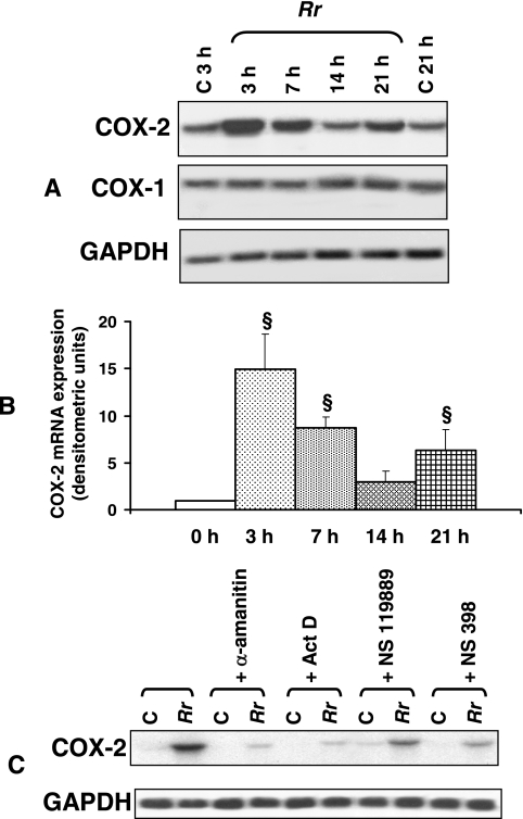
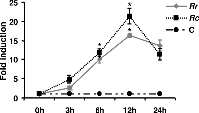
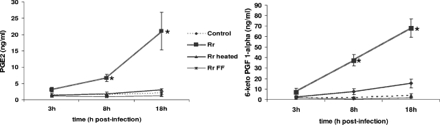
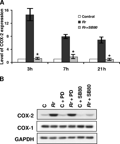
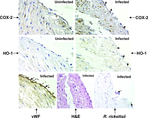
Rickettsiae, a diverse group of obligately intracellular gram-negative bacteria, include etiologic agents of the spotted fever and typhus groups of diseases. Rocky Mountain spotted fever and boutonneuse fever, due to Rickettsia rickettsii and R. conorii, respectively, are characterized by widespread infection of the vascular endothelium, microvascular injury, and vasculitis. Cultured human endothelial cells (EC) are highly susceptible to infection and respond by altering the expression of adhesion molecules, regulatory cytokines, and the antioxidant enzyme heme oxygenase (HO). In the vasculature, HO regulates the cyclooxygenase (COX) enzymes, among which the inducible isozyme COX-2 facilitates the synthesis of prostaglandins (PGs). Using in vitro and ex vivo models of infection, we demonstrate here that R. rickettsii infection of human EC causes robust induction of COX-2 mRNA and protein expression but has no apparent effect on the constitutive COX-1 isoform. Cells infected with viable rickettsiae consistently displayed significantly increased secretion of 6-keto-PGF(1alpha) and PGE(2). R. rickettsii-induced COX-2 was sensitive to inhibitors of de novo transcription and the pyridinylimidazole-based compound SB 203580, suggesting that this transcriptional host cell response involves signaling through p38 mitogen-activated protein kinase. PG production by infected cells was abrogated by NS 398 (a selective COX-2 inhibitor) and indomethacin (a pan-COX inhibitor). Immunohistochemical staining of sections of infected umbilical cords and corresponding uninfected controls revealed comparatively more intense and abundant staining for COX-2 in infected endothelia. Induction of the endothelial COX-2 system and the resultant enhanced release of vasoactive PGs may contribute to the regulation of inflammatory responses and vascular permeability changes during spotted fever rickettsioses.


Figures

FIG. 1.
FIG. 1.
Time course of R. rickettsii-induced COX-2 expression in endothelial cells. (A) Northern analysis of RNAs isolated from uninfected EC at 3 and 21 h (C, control) and from cells infected with R. rickettsii (Rr) for 3, 7, 14, and 21 h. Blots were probed in succession with 32P-labeled human-specific COX-2, COX-1, and GAPDH (housekeeping gene) cDNA probes, and the results of a typical representative experiment are shown. (B) The steady-state levels of COX-2 mRNA in R. rickettsii-infected EC at different times postinfection were normalized to that of GAPDH and compared with the average values of basal levels in simultaneously cultured cells that were left uninfected. The mean baseline COX-2 expression level in each experiment was assigned a value of 1. The results are presented as means ± standard errors for a minimum of three independent experiments, and statistically significant changes from uninfected controls (0 h) are indicated by the symbol §. (C) Effects of the transcriptional inhibitors α-amanitin (5 μg/ml) and actinomycin D (Act D; 0.25 μg/ml), the protein synthesis blocker NSC 119889 (20 μM), and the selective COX-2 inhibitor NS 398 (50 μM) on R. rickettsii-induced COX-2 mRNA expression. C, uninfected controls; Rr, R. rickettsii-infected EC.
FIG. 2.
FIG. 2.
Densitometric data obtained from immunoblot analyses using COX-2-specific antibody and normalized for loading variations by stripping and probing of the blots with a monoclonal anti-tubulin antibody (n ≥ 3) were plotted as mean levels of induction ± standard errors relative to the control (C). Rr, R. rickettsii; Rc, R. conorii. *, P ≤ 0.05.
FIG. 3.
FIG. 3.
Enhanced secretion of prostaglandins by R. rickettsii-infected EC. The levels of PGE2 and 6-keto-PGF in culture supernatants collected from uninfected controls and infected cells (Rr) at the indicated times were measured by an enzyme-linked immunosorbent assay. Also shown is a comparison of the effects of host cell interactions with viable (Rr) and heat-inactivated (Rr heated) or formalin-fixed (Rr FF) rickettsiae. Data points represent accumulated PG concentrations as means ± standard errors from at least three independent experiments. *, significant increase (P ≤ 0.05) in comparison to the baseline control level.
FIG. 4.
FIG. 4.
Inhibition of p38 MAP kinase activity decreases R. rickettsii-induced COX-2 expression. (A) Detailed densitometric analysis of COX-2 expression during R. rickettsii infection (Rr) in the presence and absence of a specific p38 MAP kinase inhibitor, SB 203580 (SB80). The results were calculated as means ± standard errors from three independent observations. *, significant reduction (P ≤ 0.05) in COX-2 mRNA level compared to that in infected cells alone, i.e., in the absence of the p38 inhibitor SB 203580. (B) Comparison of the effects of selective inhibition of ERK and p38 MAP kinase pathways via PD 98059 (PD; 10 μM) and SB 203580 (SB80; 10 μM), respectively, on the COX-2 mRNA level in uninfected (C) and R. rickettsii-infected (Rr) endothelial cells.
FIG. 5.
FIG. 5.
Immunohistochemical detection of COX-2 and HO-1 in R. rickettsii-infected endothelia from intact human umbilical veins. The serial sections of uninfected controls and infected cord specimens were stained with COX-2- and HO-1-specific antibodies. Sections from infected cords were stained for vWF and rickettsial antigen (indicated by large arrows) as described in Materials and Methods. Counterstaining with hematoxylin and eosin (H&E) was also carried out. Positive COX-2 (dark brown) and HO-1 (light brown) staining was predominantly detected in the endothelial cells of infected vasculature and is indicated by small arrows. Positive COX-2 staining in some inflammatory and mesenchymal cells of infected cord specimens was also seen. Magnification, ×400.

References

    1. Anguita, J., S. Samanta, S. K. Ananthanarayanan, B. Revilla, G. P. Geba, S. W. Barthold, and E. Fikrig. 2002. Cyclooxygenase 2 activity modulates the severity of murine Lyme arthritis. FEMS Immunol. Med. Microbiol. 34:187-191. - PMC - PubMed
    1. Appleby, S. B., A. Ristimaki, K. Neilson, K. Narko, and T. Hla. 1994. Structure of the human cyclo-oxygenase-2 gene. Biochem. J. 302:723-727. - PMC - PubMed
    1. Bach, F. H. 2005. Heme oxygenase-1: a therapeutic amplification funnel. FASEB J. 19:1216-1219. - PubMed
    1. Balsinde, J., M. A. Balboa, and E. A. Dennis. 1998. Functional coupling between secretory phospholipase A2 and cyclooxygenase-2 and its regulation by cytosolic group IV phospholipase A2. Proc. Natl. Acad. Sci. USA 95:7951-7956. - PMC - PubMed
    1. Barnett, J., J. Chow, D. Ives, M. Chiou, R. Mackenzie, E. Osen, B. Nguyen, S. Tsing, C. Bach, J. Freire, H. Chan, E. Sigal, and C. Ramesha. 1994. Purification, characterization and selective inhibition of human prostaglandin G/H synthase 1 and 2 expressed in the baculovirus system. Biochim. Biophys. Acta 1209:130-139. - PubMed

Publication types

MeSH terms