. 2006 Sep;74(9):5114-25.
doi: 10.1128/IAI.00795-06.

Characterization of the receptor-ligand pathways important for entry and survival of Francisella tularensis in human macrophages

Affiliations

Characterization of the receptor-ligand pathways important for entry and survival of Francisella tularensis in human macrophages

Ashwin Balagopal et al. Infect Immun. 2006 Sep.

Abstract

Inhalational pneumonic tularemia, caused by Francisella tularensis, is lethal in humans. F. tularensis is phagocytosed by macrophages followed by escape from phagosomes into the cytoplasm. Little is known of the phagocytic mechanisms for Francisella, particularly as they relate to the lung and alveolar macrophages. Here we examined receptors on primary human monocytes and macrophages which mediate the phagocytosis and intracellular survival of F. novicida. F. novicida association with monocyte-derived macrophages (MDM) was greater than with monocytes. Bacteria were readily ingested, as shown by electron microscopy. Bacterial association was significantly increased in fresh serum and only partially decreased in heat-inactivated serum. A role for both complement receptor 3 (CR3) and Fcgamma receptors in uptake was supported by studies using a CR3-expressing cell line and by down-modulation of Fcgamma receptors on MDM, respectively. Consistent with Fcgamma receptor involvement, antibody in nonimmune human serum was detected on the surface of Francisella. In the absence of serum opsonins, competitive inhibition of mannose receptor (MR) activity on MDM with mannan decreased the association of F. novicida and opsonization of F. novicida with lung collectin surfactant protein A (SP-A) increased bacterial association and intracellular survival. This study demonstrates that human macrophages phagocytose more Francisella than monocytes with contributions from CR3, Fcgamma receptors, the MR, and SP-A present in lung alveoli.


Figures

FIG. 1.
FIG. 1.
Cell differentiation-dependent association of F. novicida and the LVS with monocytes and MDM. (A) Monocytes or MDM were harvested from human PBMC at either day 1 or day 5 to 6, respectively, adhered to glass coverslips, and incubated for 2 h with F. novicida at an MOI of 10:1 or the LVS at an MOI of 60:1 in the absence or presence of increasing concentrations of autologous serum. Cell association was quantified by counting F. novicida cells associated with 150 to 200 consecutive cells. Data are the means ± standard deviations of bacteria per cell (triplicate wells) in representative experiments for F. novicida (n = 3) and the LVS (n = 2). Paired one-tail Student t tests were used to generate P values. (B) MDM monolayers on glass coverslips were incubated with either GFP-expressing F. novicida or LVS for 2 h, and bacteria per macrophage were enumerated using fluorescence microscopy. Results were normalized to the mean number of F. novicida cells per macrophage in both the “no-serum” and “serum” groups. The results shown are the means ± SEM from six independent experiments, three at an MOI of 10:1 and three at an MOI of 600:1. *, P = 0.0163; **, P = 0.0016 (paired two-tailed Student t test). (C) MDM monolayers were incubated with F. novicida or the LVS for 2 h and then incubated with gentamicin and lysed in 0.1% Triton X-100, and MDM lysates were plated on chocolate agar for CFU. Results shown are the means ± SEM from two independent experiments. *, P = 0.0194; **, P = 0.0023 (unpaired Student t test).
FIG. 2.
FIG. 2.
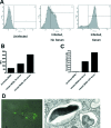
Serum-dependent cell association of F. novicida with MDM. (A to C) GFP-expressing F. novicida-infected MDM were analyzed by flow cytometry. Uninfected MDM were used to set the gates. Shown is an analysis of histograms (A) and bar graphs showing the mean fluorescence intensities (MFI) (B) and percentages of positive cells infected (C) of uninfected MDM and GFP-expressing F. novicida-infected MDM in the absence and presence of 2.5% serum. Data shown are representative of two experiments. (D, left panel) Following incubation of MDM for 2 h, nonadherent F. novicida cells were washed away, and MDM on coverslips were permeabilized, washed, and then incubated with mouse anti-F. novicida antibody followed by Alexa Fluor 488-conjugated goat anti-mouse IgG. Bacterial association was visualized by indirect immunofluorescence microscopy. Shown is a representative image of an overlay of light- and fluorescence-microscopic images of an MDM with associated fluorescent F. novicida (magnification, ×1,000). (D, right panel) After a 2-h incubation, MDM were fixed and prepared for transmission EM. A representative cross section showing ingested F. novicida cells contained within phagosomal membranes is shown (magnification, ×28,000).
FIG. 3.
FIG. 3.
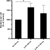
Heat-labile and stable components of serum contribute to Francisella association with MDM. GFP-expressing F. novicida or wild-type F. novicida cells were opsonized in media alone, 2.5% serum, or 2.5% HI serum for 30 min at 37°C. Bacteria were then added to MDM monolayers for 2 h at an MOI of 50:1. Cells were fixed and, in the case of wild-type F. novicida, permeabilized and immunostained, and the bacteria per MDM were enumerated by fluorescence microscopy. One hundred fifty to 200 consecutive MDM on each coverslip of triplicate coverslips/test group were counted. Results shown are the means ± SEM from three independent experiments. *, P<0.05 (paired one-tailed Student t test).
FIG. 4.
FIG. 4.
CR3 is important in the association of Francisella. Wild-type CHO and CHO-CR3 cells were adhered to glass coverslips overnight and then incubated with F. novicida cells that had been preopsonized in media alone, 10% fresh serum, or 10% HI serum at MOIs of 100:1 and 50:1. After fixation, permeabilization, and immunostaining, >500 consecutive cells per coverslip were counted and enumerated by immunofluorescence microscopy. Results shown are means ± SEM from two independent experiments. *, P < 0.05 (unpaired two-tailed Student t test).
FIG. 5.
FIG. 5.
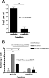
FcγR on human macrophages mediate the association of Francisella. MDM were adhered to coverslips that had been coated with HSA (control) or HSA and rabbit anti-HSA antibody to down-modulate FcγR. (A) E-IgG were incubated with control or FcγR-down-modulated MDM for 1 h at an MOI of 10:1. After fixation, E-IgG per MDM were counted by light microscopy. A representative experiment is shown (means ± standard deviations [SD] of triplicate wells). **, P = 0.0025 (paired two-tailed Student t test). (B) GFP-expressing F. novicida cells were preopsonized in media alone, 10% fresh serum, or 10% HI serum and added to control or FcγR-down-modulated MDM for 2 h at an MOI of 500:1. After fixation, GFP-expressing F. novicida cells per MDM were counted by fluorescence microscopy. Results shown are from one representative experiment (n = 4). Means ± SD of triplicate wells are shown. *, P < 0.01; **, P < 0.001 (calculated by two-way analysis of variance comparing multiple samples and Bonferroni posttests). External comparisons, e.g., “10% serum, −antiHSA” versus “10% serum, +antiHSA,” were made using a two-tailed Student t test, with P values as indicated.
FIG. 6.
FIG. 6.
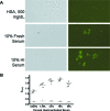
Antibody in nonimmune human serum recognizes F. novicida. F. novicida cells (1 × 10⁸) were incubated with various concentrations of fresh nonimmune human serum, HI serum, or HSA control for 30 min at 37°C, washed, and blocked with 3% ovalbumin. (A) Triplicate samples were then incubated with FITC-conjugated anti-human IgG, IgA, or IgM antibody, washed, and placed on slides. The presence or absence of fluorescent bacteria was assessed by immunofluorescence microscopy. Approximately 10 fields per coverslip were examined in each group. The photomicrographs shown are representative high-power fields (×1,000). (B) Serum- or HSA-treated samples (1 × 10⁷ bacteria, in triplicate) were dried in ELISA plate wells overnight. Wells were then blocked, washed, and incubated with HRP-conjugated anti-human IgG, IgA, or IgM antibody. Subsequently, wells were incubated with HRP substrate and reactions were terminated with 2% oxalic acid. A415 was measured. The results from six or seven donors, one immune donor (arrows) and five or six nonimmune donors, are shown. Each dot represents the mean of triplicate wells for each donor. The solid horizontal lines are the means for donors, and the dashed horizontal lines are the means for the HSA groups. *, P < 0.05; **, P < 0.005; ***, P < 0.0005 (paired two-tailed Student t test for HSA relative to the serum groups). The immune donor's result was excluded from the statistical comparisons.
FIG. 7.
FIG. 7.
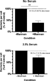
The macrophage MR mediates nonopsonic association of F. novicida. MDM monolayers on glass coverslips were preincubated with either soluble mannan or media alone for 30 min at 37°C in 5% CO2 and then incubated with GFP-expressing F. novicida at an MOI of 600:1 for 2 h. After fixing, F. novicida cells per MDM were enumerated using fluorescence microscopy. Infections were performed in the absence (A) and presence (B) of 2.5% autologous serum. Results from four independent experiments are shown (means ± SEM). **, P = 0.0074 (paired two-tailed Student t test).
FIG. 8.
FIG. 8.
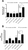
Preopsonization of F. novicida with SP-A increases the association of bacteria with MDM. MDM monolayers on glass coverslips were incubated without (control) or with SP-A before or at the time of infection with GFP-expressing F. novicida at an MOI of 600:1. As indicated, some MDM monolayers were washed prior to adding bacteria. After 2 h and a washing, MDM were fixed and bacteria per MDM were enumerated using fluorescence microscopy (A, left four bars). Results shown are the means ± SEM normalized to the “no-SP-A” group (n = 2). (A, right two bars) GFP-expressing F. novicida cells were preopsonized with or without SP-A and added to MDM. After 2 h and a washing, MDM were fixed and bacteria per MDM were enumerated using fluorescence microscopy. Results shown are the means ± SEM normalized to the “mock-opsonization” group (n = 3). *, P = 0.0107 (paired two-tailed Student t test). (B) SP-A treatment of MDM and bacteria was identical to that for panel A. After 2 h and a washing, extracellular bacteria were killed with gentamicin and CFU of MDM lysates were obtained. Results shown are the means ± SEM from three independent experiments.

References

    1. Abd, H., T. Johansson, I. Golovliov, G. Sandstrom, and M. Forsman. 2003. Survival and growth of Francisella tularensis in Acanthamoeba castellanii. Appl. Environ. Microbiol. 69:600-606. - PMC - PubMed
    1. Allavena, P., M. Chieppa, P. Monti, and L. Piemonti. 2004. From pattern recognition receptor to regulator of homeostasis: the double-faced macrophage mannose receptor. Crit. Rev. Immunol. 24:179-192. - PubMed
    1. Alluisi, E. A., W. R. Beisel, P. J. Bartelloni, and G. D. Coates. 1973. Behavioral effects of tularemia and sandfly fever in man. J. Infect. Dis. 128:710-717. - PubMed
    1. Anthony, L. D., R. D. Burke, and F. E. Nano. 1991. Growth of Francisella spp. in rodent macrophages. Infect. Immun. 59:3291-3296. - PMC - PubMed
    1. Beharka, A. A., C. D. Gaynor, B. K. Kang, D. R. Voelker, F. X. McCormack, and L. S. Schlesinger. 2002. Pulmonary surfactant protein A up-regulates activity of the mannose receptor, a pattern recognition receptor expressed on human macrophages. J. Immunol. 169:3565-3573. - PubMed

Publication types

MeSH terms

LinkOut - more resources