. 2006 Sep;74(9):5211-20.
doi: 10.1128/IAI.00056-06.

Actinobacillus actinomycetemcomitans outer membrane protein 100 triggers innate immunity and production of beta-defensin and the 18-kilodalton cationic antimicrobial protein through the fibronectin-integrin pathway in human gingival epithelial cells

Affiliations

Actinobacillus actinomycetemcomitans outer membrane protein 100 triggers innate immunity and production of beta-defensin and the 18-kilodalton cationic antimicrobial protein through the fibronectin-integrin pathway in human gingival epithelial cells

Kazuhisa Ouhara et al. Infect Immun. 2006 Sep.

Abstract

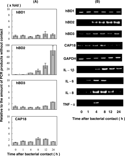
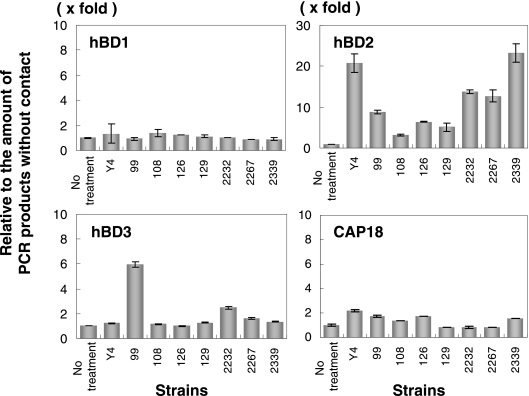
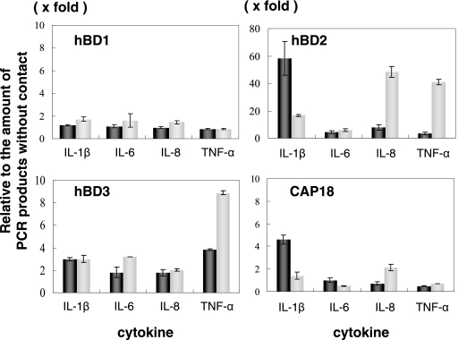
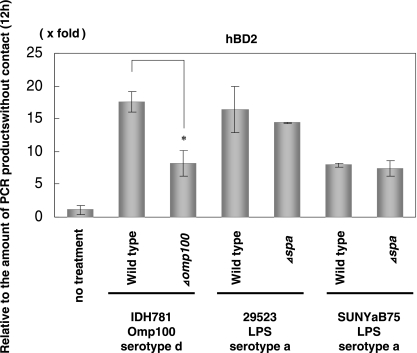
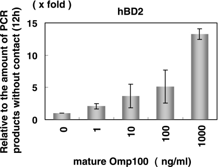
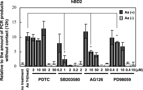
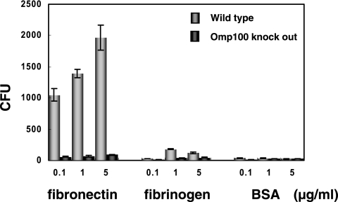
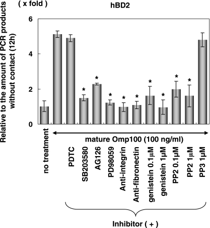
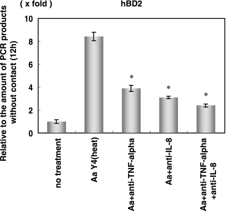
Antimicrobial peptides, human beta-defensin (hBD), and the 18-kDa cationic antimicrobial protein (CAP18) are components of innate immunity. These peptides have antimicrobial activity against bacteria, fungi, and viruses. Actinobacillus actinomycetemcomitans is a gram-negative facultative anaerobe implicated in the initiation of periodontitis. The innate immunity peptides have antibacterial activity against A. actinomycetemcomitans. We investigated the molecular mechanism of human gingival epithelial cells (HGEC) responding to exposure to A. actinomycetemcomitans. HGEC constitutively express hBD1 and inducibly express hBD2, hBD3, and CAP18 on exposure to A. actinomycetemcomitans. The level of expression varies among clinical isolates. In the signaling pathway for hBD2 induction by the bacterial contact, we demonstrate that the mitogen-activated protein (MAP) kinase and not the NF-kappaB transcription factor pathway is used. We found the outer membrane protein 100 (Omp100; identified by molecular mass) is the component inducing the hBD2 response. Omp100 binds to fibronectin, an extracellular matrix inducing hBD2 via the MAP kinase pathway. Anti-integrin alpha(5)beta(1), antifibronectin, genistein, and PP2 suppress the Omp100-induced expression of hBD2, suggesting that Src kinase is involved through integrin alpha(5)beta(1). The inflammatory cytokines, tumor necrosis factor alpha (TNF-alpha), interleukin-1beta (IL-1beta), IL-6 and IL-8, produced by HGEC on contact with A. actinomycetemcomitans also stimulate expression of hBD2. Further, neutralizing antibody against TNF-alpha or IL-8 partially inhibits the induction of hBD2 on bacterial contact. Therefore, we found that the induction of the antimicrobial peptides is mediated by a direct response principally through an Omp100-fibronectin interaction, and using secondary stimulation by inflammatory cytokines induced by the bacterial exposure.


Figures

FIG. 1.
FIG. 1.
Expression of β-defensins, CAP18, and cytokine mRNA in HGEC stimulated with heat-inactivated A. actinomycetemcomitans Y4 cells. The total RNA was extracted from cells after exposure to A. actinomycetemcomitans at various times and then used for real-time PCR (A) and RT-PCR (B) performed as described in Materials and Methods. The results of the real-time PCR are expressed as a ratio comparing the value at time zero to the value at the data time point. The values represent the means ± standard deviations of triplicate experiments.
FIG. 2.
FIG. 2.
Expression of hBD1, hBD2, hBD3, and CAP18 mRNA in HGEC stimulated with heat-inactivated clinically isolated A. actinomycetemcomitans cells. The total RNA was extracted from cells after exposure to A. actinomycetemcomitans for 12 h and used for real-time PCR performed as described in Materials and Methods. The results of the real-time PCR are expressed as a ratio comparing the value at time zero to the value at the data time point. The values represent the means ± standard deviations of triplicate experiments.
FIG. 3.
FIG. 3.
Expression of hBD1, hBD2, hBD3, and CAP18 mRNA in HGEC stimulated with IL-1β, IL-6, IL-8, and TNF-α. The total RNA was extracted from the cells after the addition of several cytokines for 12 h and then used for real-time PCR performed as described in Materials and Methods. Black bars, 1 ng/ml; gray bars, 10 ng/ml. The results of the real-time PCR are expressed as a ratio comparing the value at time zero to the value at the data time point. The values represent the means ± standard deviations of triplicate experiments.
FIG. 4.
FIG. 4.
Effect of Omp100 and SPA on the expression of hDB2. Heat-inactivated A. actinomycetemcomitans strains were added to HGEC and incubated for 12 h. The total RNA was extracted from cells after contact with bacteria for 12 h and used for real-time PCR. The results of the real-time PCR are expressed as a ratio comparing the value at time zero to the value at the data time point. The values represent the means and ± standard deviations of triplicate experiments.
FIG. 5.
FIG. 5.
Effect of purified A. actinomycetemcomitans Y4 Omp100 on the expression of hDB2 in HGEC. Purified Omp100 was incubated with HGEC at various concentrations for 12 h. After the total RNA extraction, real-time PCR was performed. The results of the real-time PCR are expressed as a ratio comparing the value at time zero to the value at the data time point. The values represent the means ± standard deviations of triplicate experiments.
FIG. 6.
FIG. 6.
Effect of inhibitors on hBD2 mRNA induction by A. actinomycetemcomitans cells. HGEC were pretreated with PDTC (NF-κB inhibitor; 2 to 50 μM), SB203580 (p38 inhibitor; 0.2 to 5 μM), AG126 (JNK inhibitor, 2 to 50 μM), or PD98059 (ERK inhibitor; 0.4 to 10 μM) for 1 h and then stimulated with heat-inactivated A. actinomycetemcomitans Y4 for 12 h. The total RNA was extracted and analyzed using real-time PCR. Gray bar, without bacterial contact; black bar, with bacterial contact. The results of the real-time PCR are expressed as a ratio in comparison to the value at time zero, and the values represent the means ± standard deviations of triplicate cultures. *, differs significantly (P < 0.01) from cells stimulated with A. actinomycetemcomitans Y4 alone.
FIG. 7.
FIG. 7.
Binding assay of A. actinomycetemcomitans strains expressing Omp100 and knockout mutant. The binding assay was performed as described in Materials and Methods. Gray bar, wild type; black bar, Omp100 knockout mutant. Values represent the means ± standard deviations of triplicate wells.
FIG. 8.
FIG. 8.
Effect of inhibitors on hBD2 mRNA expression by Omp100. HGEC were pretreated with PDTC (NF-κB inhibitor; 2 μM), SB203580 (p38 inhibitor; 0.2 μM), AG126 (JNK inhibitor; 2 μM), PD98059 (ERK inhibitor; 0.4 μM), anti-human fibronectin affinity-isolated antibody, anti-integrin affinity-isolated antibody, PP2 (selective Src tyrosine kinase inhibitor), PP3 (negative control for the Src tyrosine kinase inhibitor), and genistein (protein tyrosine kinases inhibitor) with each concentration for 1 h and then stimulated with purified Omp100 (100 ng/ml) for 12 h. The total RNA was extracted and analyzed using real-time PCR. The results of the real-time PCR are expressed as a ratio comparing the value at time zero to the value at the data time point. The values represent the means and ± standard deviations of triplicate experiments. *, differs significantly (P < 0.01) from cells stimulated with Omp100 alone.
FIG. 9.
FIG. 9.
Effect of anti-TNF-α and anti-IL-8 on hBD2 mRNA induction. HGEC were pretreated with monoclonal anti-human TNF-α antibody and monoclonal anti-human IL-8 antibody (final concentration, 100 ng/ml) for 1 h and then stimulated with heat-inactivated A. actinomycetemcomitans Y4 for 12 h. The total RNA was extracted and analyzed using real-time PCR. The results of the real-time PCR are expressed as a ratio comparing the value at time zero to the value at the data time point. The values represent the means and ± standard deviations of triplicate experiments. *, differs significantly (P < 0.01) from cells stimulated with A. actinomycetemcomitans Y4 alone.

References

    1. Agerer, F., S. Lux, M. A., M. Rohde, K. Ohlsen, and C. R. Hauck. 2005. Cellular invasion by Staphylococcus aureus reveals a functional link between focal adhesion kinase and cortactin in integrin-mediated internalization. J. Cell Sci. 118:2189-2200. - PubMed
    1. Asakawa, R., H. Komatsuzawa, T. Kawai, S. Yamada, R. B. Goncalves, S. Izumi, T. Fujiwara, Y. Nakano, N. Suzuki, Y. Uchida, K. Ouhara, H. Shiba, M. A. Taubman, H. Kurihara, and M. Sugai. 2003. Outer membrane protein 100, a versatile virulence factor of Actinobacillus actinomycetemcomitans. Mol. Microbiol. 50:1125-1139. - PubMed
    1. Asikainen, S., C. Chen, M. Saarela, L. Saxen, and J. Slots. 1997. Clonal specificity of Actinobacillus actinomycetemcomitans in destructive periodontal disease. Clin. Infect. Dis. 25(Suppl. 2):S227-S229. - PubMed
    1. Bals, R. 2000. Epithelial antimicrobial peptides in host defense against infection. Respir. Res. 1:141-150. - PMC - PubMed
    1. Bals, R., X. Wang, Z. Wu, T. Freeman, V. Bafna, M. Zasloff, and J. M. Wilson. 1998. Human beta-defensin 2 is a salt-sensitive peptide antibiotic expressed in human lung. J. Clin. Investig. 102:874-880. - PMC - PubMed

Publication types

MeSH terms

LinkOut - more resources