. 2006 Sep;74(9):5292-301.
doi: 10.1128/IAI.02024-05.

Intestinal intraepithelial lymphocytes sustain the epithelial barrier function against Eimeria vermiformis infection

Affiliations

Intestinal intraepithelial lymphocytes sustain the epithelial barrier function against Eimeria vermiformis infection

Kyoko Inagaki-Ohara et al. Infect Immun. 2006 Sep.

Abstract

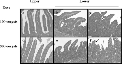
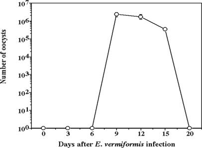
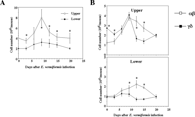
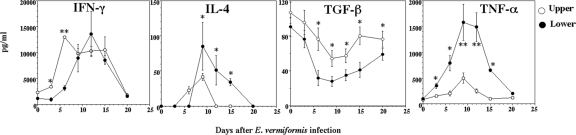
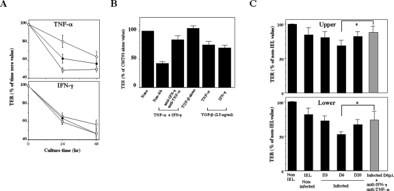
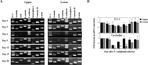
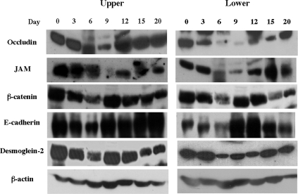
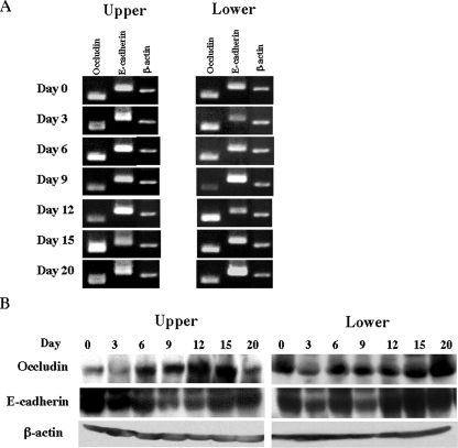
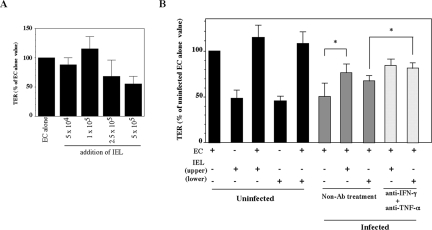
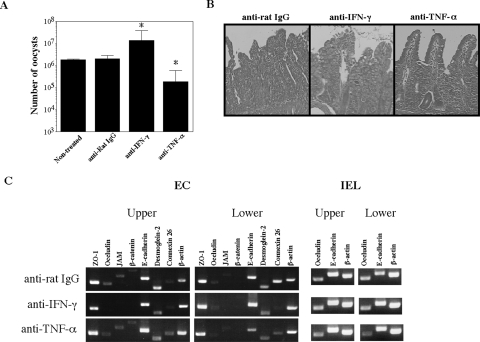
Eimeria spp. are intracellular protozoa that infect intestinal epithelia of most vertebrates, causing coccidiosis. Intestinal intraepithelial lymphocytes (IEL) that reside at the basolateral site of epithelial cells (EC) have immunoregulatory and immunoprotective roles against Eimeria spp. infection. However, it remains unknown how IEL are involved in the regulation of epithelial barrier during Eimeria sp. infection. Here, we demonstrated two distinct roles of IEL against infection with Eimeria vermiformis, a murine pathogen: production of cytokines to induce protective immunity and expression of junctional molecules to preserve epithelial barrier. The number of IEL markedly increased when oocyst production reached a peak. During infection, IEL increased production of gamma interferon (IFN-gamma) and tumor necrosis factor alpha (TNF-alpha) and decreased transforming growth factor beta (TGF-beta) production. Addition of IFN-gamma and TNF-alpha or supernatants obtained from cultured IEL from E. vermiformis-infected mice reduced transepithelial electrical resistance (TER) in a confluent CMT93 cell monolayer, a murine intestine-derived epithelial line, but antibodies against these cytokines suppressed the decline of TER. Moreover, TGF-beta attenuated the damage of epithelial monolayer and changes in TER caused by IFN-gamma and TNF-alpha. The expression of junctional molecules by EC was decreased when IEL produced a high level of IFN-gamma and TNF-alpha and a low level of TGF-beta in E. vermiformis-infected mice. Interestingly, IEL constantly expressed junctional molecules and a coculture of EC with IEL increased TER. These results suggest that IEL play important multifunctional roles not only in protection of the epithelium against E. vermiformis-induced change by cytokine production but also in direct interaction with the epithelial barrier when intra-EC junctions are down-regulated.


Figures

FIG. 1.
FIG. 1.
Hematoxylin-and-eosin-stained transverse sections of the small intestine on day 9 p.i. Mice were given 100 or 500 oocysts of E. vermiformis. On day 9 p.i., the small intestine was removed and divided into parts, the upper (including duodenum [a and d]) and the lower (jejunum [b and e] and ileum [c and f]) (magnification, ×200).
FIG. 2.
FIG. 2.
Oocyst output of E. vermiformis in C57BL/6 mice. Mice were orally infected with 100 oocysts of E. vermiformis, and the oocyst output was determined every 3 days. Three mice were used in each point. Values represent the mean ± standard deviation of three individual experiments.
FIG. 3.
FIG. 3.
Cell number of IEL following infection with E. vermiformis. (A) Total IEL number was counted by staining with trypan blue. (B) Population of IEL bearing TCRαβ and TCRγδ of the upper and lower parts following infection. IEL were stained with fluorescein isothiocyanate-conjugated TCRγδ MAb, PE-conjugated TCRβ MAb, and allophycocyanin-conjugated CD3ɛ MAb. The TCRαβ and TCRγδ expression was gated on CD3+ cells. Three mice were used in each point. Values represent the mean ± standard deviation of three individual experiments in a triplicate assay. Asterisks represent statistically significant differences (P < 0.05).
FIG. 4.
FIG. 4.
Changes in cytokine production of IEL following infection with E. vermiformis. Freshly isolated IEL at each time point were cultured with plate-bound anti-CD3 MAb for 48 h, and the supernatants were collected to determine the concentration of each cytokine by ELISA for IFN-γ, IL-4, TNF-α, and TGF-β. Three mice were used in each point. Values represent the mean ± standard deviation of three individual experiments in a triplicate assay. Asterisks represent statistically significant differences (*, P < 0.05; **, P < 0.01).
FIG. 5.
FIG. 5.
Abolishment of TER in an epithelial cell line by inflammatory cytokines produced by IEL. (A) CMT93 cells were exposed to 1 (open circles), 10 (closed circles), and 100 (open squares) ng/ml of recombinant IFN-γ or TNF-α for 24 and 48 h. (B) CMT93 cells were cultured in combination with 10 ng/ml of IFN-γ and TNF-α. for 48 h. CMT93 cells were also cultured with 2.5 ng/ml of TGF-β1 and IFN-γ or TNF-α (10 ng/ml) for 48 h. (C) CMT93 cells were cultured with supernatants of IEL prepared from mice infected with E. vermiformis on days 3, 6, and 20 p.i. in the presence of immobilized anti-CD3 MAb. Supernatant prepared from mice infected with E. vermiformis on day 6 p.i. was added with 10 μg/ml of anti-IFN-γ and anti-TNF-α Abs to CMT93 cells. These cells were then incubated for 48 h at 37°C for the determination of TER. Values represent the mean ± standard deviation of three individual experiments in a triplicate assay. Asterisks represent statistically significant differences (*, P < 0.05).
FIG. 6.
FIG. 6.
Decreased mRNA expression of junctional molecules in small intestinal EC following infection with E. vermiformis. (A) Total RNA was prepared from EC of small intestine and amplified by RT-PCR. (B) mRNA expression of ZO-1 and occludin was analyzed quantitatively by real-time PCR. Data are presented as the ratio of each time point to day 0 in the upper and lower segment. Asterisks represent statistically significant differences between the upper and lower segments (*, P < 0.05; **, P < 0.01).
FIG. 7.
FIG. 7.
Western blotting analysis for the expression of junctional molecules at the protein level in EC of small intestine. Cell lysate was prepared from small intestinal EC in mice and analyzed by Western blotting as described in Materials and Methods.
FIG. 8.
FIG. 8.
Sustained mRNA expression of junctional molecules in small intestinal IEL following infection with E. vermiformis. After small intestine was separated into upper and lower segments, fresh IEL were obtained. (A) Total RNA was prepared from IEL in the upper and lower parts and amplified by RT-PCR. (B) Western blotting analysis for the expression of junctional molecules at protein level in IEL of small intestine. Cell lysate was prepared from small intestinal IEL in the upper and lower segments and analyzed by Western blotting.
FIG. 9.
FIG. 9.
Increased TER in freshly isolated EC cocultured with IEL from E. vermiformis-infected mice on day 6 p.i. (A) Effect of a varying number of IEL on TER of EC. On day 4 after EC were cultured, IEL were added to EC and TER was measured on day 6. Values represent the mean ± standard deviation of three individual experiments in a triplicate assay. (B) On day 4 after EC were cultured, 1 × 10⁵ IEL were added to EC, and TER was measured with or without 10 μg/ml of anti-IFN-γ and anti-TNF-α Abs on day 6. Values represent the mean ± standard deviation of three individual experiments in a triplicate assay. Asterisks represent statistically significant differences (P < 0.05).
FIG. 10.
FIG. 10.
Modulation of junctional molecules due to depletion of IFN-γ or TNF-α in vivo. (A) Oocyst output of E. vermiformis in mice treated with anti-IFN-γ Ab or anti-TNF-α Ab in vivo. Mice were orally infected with 100 oocysts of E. vermiformis, and the oocyst output was determined on day 9 p.i. Values represent the mean ± standard deviation of three mice in each group. Asterisks represent statistically significant differences (*, P < 0.05). (B) Hematoxylin-and-eosin-stained transverse sections of the small intestine on day 9 p.i. (magnification, ×200). (C) mRNA expression of junctional molecules in small intestinal EC following infection with E. vermiformis. Total RNA was prepared from EC of small intestine and amplified by RT-PCR. IgG, immunoglobulin G.

Similar articles

Cited by

References

    1. Baumgart, D. C., W. A. Olivier, T. Reya, D. Peritt, J. L. Rombeau, and S. R. Carding. 1998. Mechanisms of intestinal epithelial cell injury and colitis in interleukin 2 (IL2)-deficient mice. Cell. Immunol. 187:52-66. - PubMed
    1. Berkes, J., V. K. Viswanathan, S. D. Savkovic, and G. Hecht. 2003. Intestinal epithelial responses to enteric pathogens: effects on the tight junction barrier, ion transport, and inflammation. Gut 52:439-451. - PMC - PubMed
    1. Blagburn, B. L., and K. S. Todd, Jr. 1984. Pathological changes and immunity associated with experimental Eimeria vermiformis infections in Mus musculus. J. Protozool. 31:556-561. - PubMed
    1. Chapman, H. D. 2001. Use of anticoccidial drugs in broiler chickens in the USA: analysis for the years 1995 to 1999. Poult. Sci. 80:572-580. - PubMed
    1. Chobotar, B., H. D. Danforth, and R. Entzeroth. 1993. Ultrastructural observations of host-cell invasion by sporozoites of Eimeria papillata in vivo. Parasitol. Res. 79:15-23. - PubMed

Publication types

MeSH terms

LinkOut - more resources