

doi: 10.1128/IAI.00332-06.
Pulmonary lymphatics are primary sites of Mycobacterium tuberculosis infection in guinea pigs infected by aerosol
Affiliations
- PMID: 16926435
- PMCID: PMC1594862
- DOI: 10.1128/IAI.00332-06
Item in Clipboard
Pulmonary lymphatics are primary sites of Mycobacterium tuberculosis infection in guinea pigs infected by aerosol
Infect Immun.
2006 Sep.
Abstract
Mycobacterium tuberculosis causes a lymphatic vasculitis in the lungs of guinea pigs infected by a low-dose aerosol. This observation suggests that in addition to being a direct conduit from the lungs to the regional lymph nodes, pulmonary lymphatics are themselves sites of infection and could be the site of latent infection.
Figures
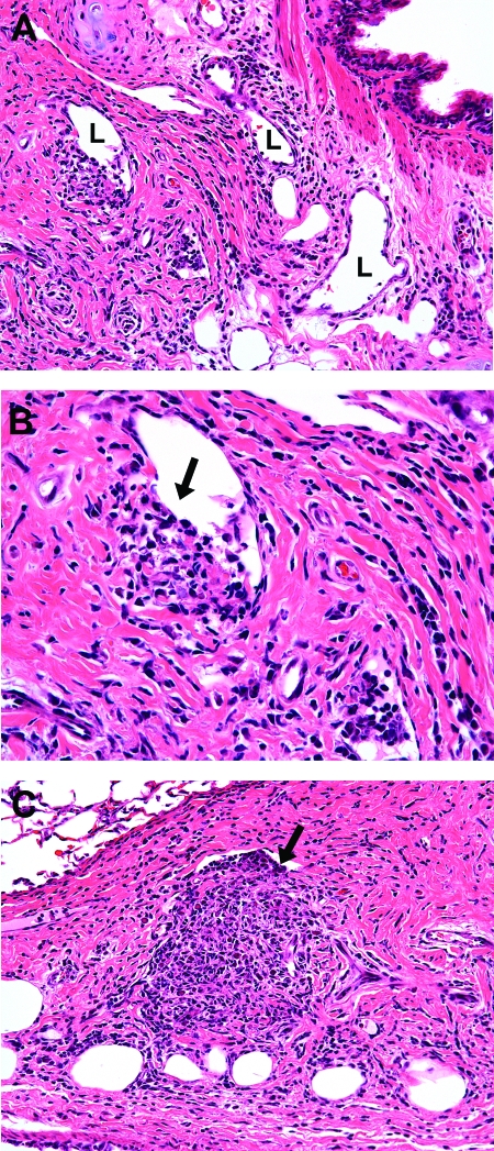
Photomicrographs of lungs of guinea pigs at 15 days postinfection with the H37Rv strain of M. tuberculosis. (A) Mixed inflammatory cells, including lymphocytes, macrophages, and granulocytes (heterophils and eosinophils), are present in the peribronchial connective tissue, which also contains variably sized lymphatic (L) vessels (magnification, ×200). (B) Small aggregates of inflammatory cells are present in the immediate perilymphatic connective tissue (arrow) and communicate with the lumen, which is partially devoid of endothelium (magnification, ×400). (C) A similar but larger accumulation of mixed inflammatory cells is present in the perilymphatic connective tissue that extends into the lymphatic lumen (arrow) (magnification, ×400). Tissues were stained with H&E.
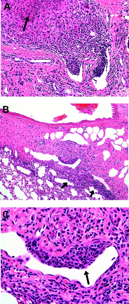
Photomicrographs of lungs of guinea pigs at 20 days postinfection with the H37Rv strain of M. tuberculosis. (A) Mixed inflammatory cells, including lymphocytes, macrophages, and granulocytes (heterophils and eosinophils), are present in the perilymphatic connective tissue, with early intralesional necrosis (arrow) and granulocyte infiltration (magnification, ×200). (B) Mixed inflammation extends from the periarterial and perilymphatic connective tissue and into the surrounding (arrows) pulmonary parenchyma (magnification, ×100). (C) Higher magnification of the field shown in panel B, demonstrating lymphangitis where the mixed inflammation infiltrates the wall (arrow) of a lymphatic vessel (magnification, ×400). Tissues were stained with H&E.
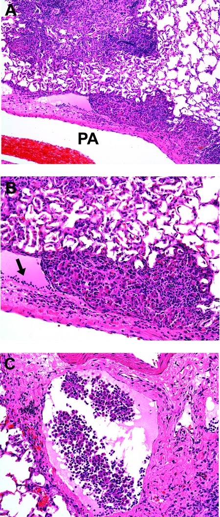
Photomicrographs of lungs of guinea pigs at 30 days postinfection with the H37Rv strain of M. tuberculosis. (A) Longitudinal section through a branch of a pulmonary artery (PA), showing parallel, nodular infiltrates of mixed inflammatory cells within the lumen of a periarterial lymphatic (lymphangitis), which is shown at a higher magnification in panel B (magnification, ×100). (B) Inflammatory cells occlude the lumen of a periarterial lymphatic that is distended with protein-rich acidophilic fluid and marginating (arrow) lymphocytes (magnification, ×400). (C) Markedly dilated periarterial lymphatic that contains circulating mixed inflammatory cells with no evidence of lymphangitis (magnification, ×200). Tissues were stained with H&E.
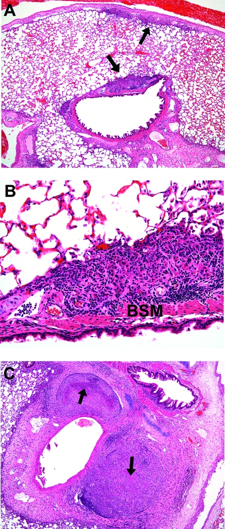
Photomicrographs of lungs of guinea pigs at 30 days postinfection with the H37Rv strain of M. tuberculosis. (A) Mixed inflammatory cells infiltrate the periarterial (small arrow) and peribronchial (large arrow) connective tissue, which is shown at a higher magnification in panel B (magnification, ×40). (B) Peribronchial mixed inflammation within the peribronchial lymphatics deep to the bronchial smooth muscle (BSM). There is minimal extension of inflammation into the surrounding alveoli of the pulmonary parenchyma (magnification, ×200). (C) Multiple well-organized lesions in the periarterial connective tissue of an immature animal. Margins are often sharply demarcated from the more loosely organized connective tissue stroma than that in mature animals. Lesions have extensive, coalescing foci (arrows) of central necrosis (magnification, ×40). Tissues were stained with H&E.
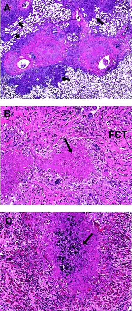
Photomicrographs of lungs of guinea pigs at 60 days postinfection with the H37Rv strain of M. tuberculosis. (A) Multiple lesions in the periarterial connective tissue coalesce within the connective tissue stroma and are continuous with inflammatory lesions that extend into the surrounding alveoli (arrows) of the pulmonary parenchyma (magnification, ×20). (B) Higher-magnification image of that shown in panel A, showing central necrosis (arrow) with resolving inflammation which is replaced by fibrous connective tissue (FCT) (magnification, ×200). (C) More advanced periarterial lesions show evidence of early mineralization (arrow) of the central necrosis (magnification, ×200). Tissues were stained with H&E.
References
-
- Abadie, V., E. Badell, P. Douillard, D. Ensergueix, P. J. Leenen, M. Tanguy, L. Fiette, S. Saeland, B. Gicquel, and N. Winter. 2005. Neutrophils rapidly migrate via lymphatics after Mycobacterium bovis BCG intradermal vaccination and shuttle live bacilli to the draining lymph nodes. Blood 106:1843-1850. - PubMed
-
- Basaraba, R. J., D. D. Dailey, C. T. McFarland, C. A. Shanley, E. E. Smith, D. N. McMurray, and I. M. Orme. 2006. Lymphadenitis as a major element of disease in the guinea pig model of tuberculosis. Tuberculosis (Edinburgh) [Epub ahead of print.] - PubMed
-
- Dheda, K., H. Booth, J. F. Huggett, M. A. Johnson, A. Zumla, and G. A. Rook. 2005. Lung remodeling in pulmonary tuberculosis. J. Infect. Dis. 192:1201-1209. - PubMed
Publication types
MeSH terms
Substances
Grants and funding
LinkOut - more resources
Full Text Sources
Other Literature Sources
