. 2006 Oct 20;498(6):810-20.
doi: 10.1002/cne.21089.

Distinct perisynaptic and synaptic localization of NMDA and AMPA receptors on ganglion cells in rat retina

Affiliations

Distinct perisynaptic and synaptic localization of NMDA and AMPA receptors on ganglion cells in rat retina

Jun Zhang et al. J Comp Neurol. .

Abstract

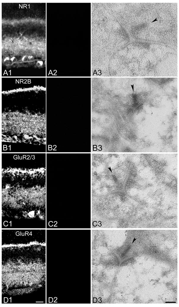
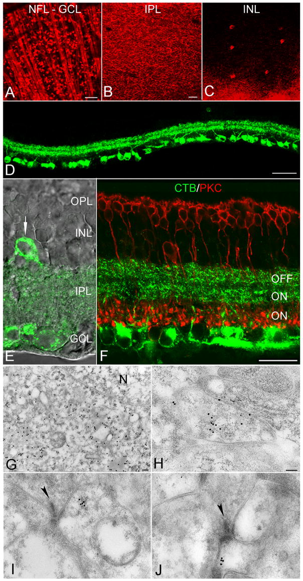
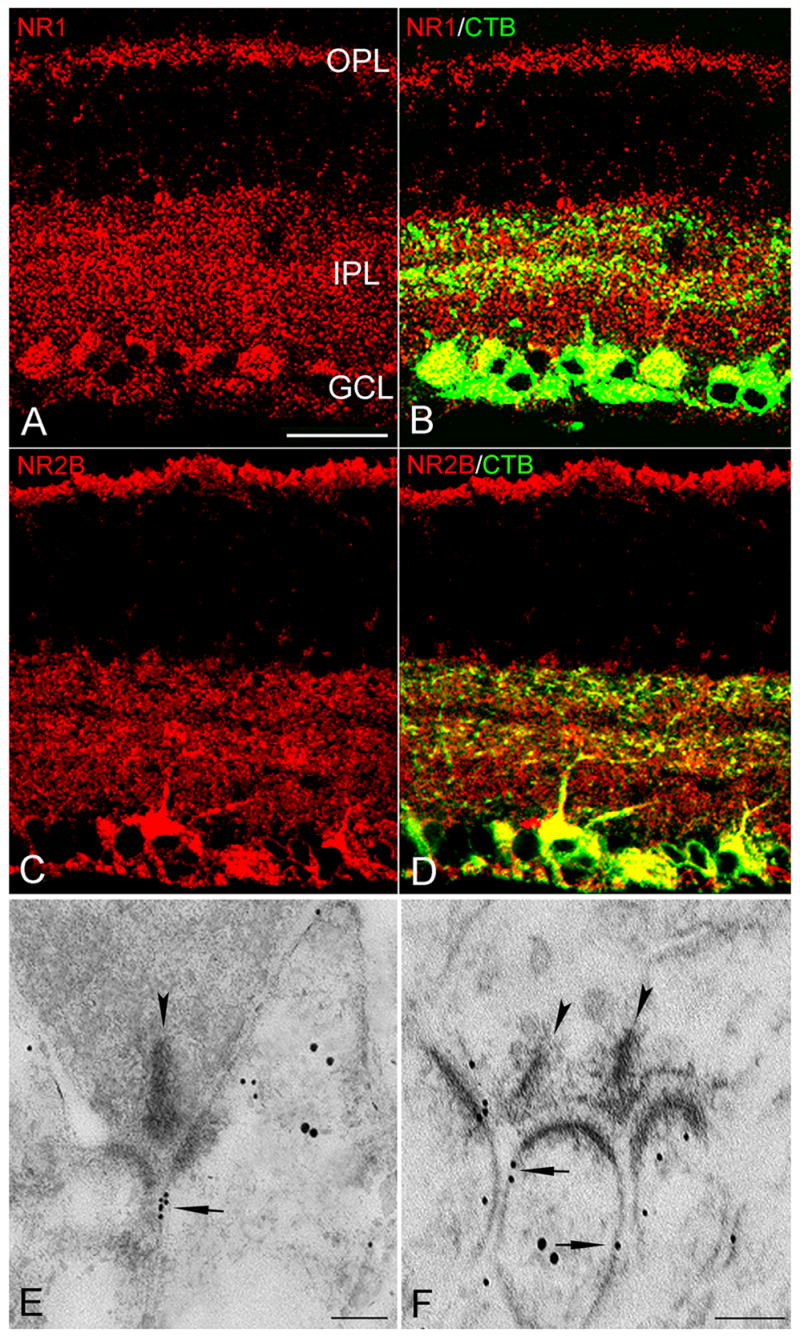
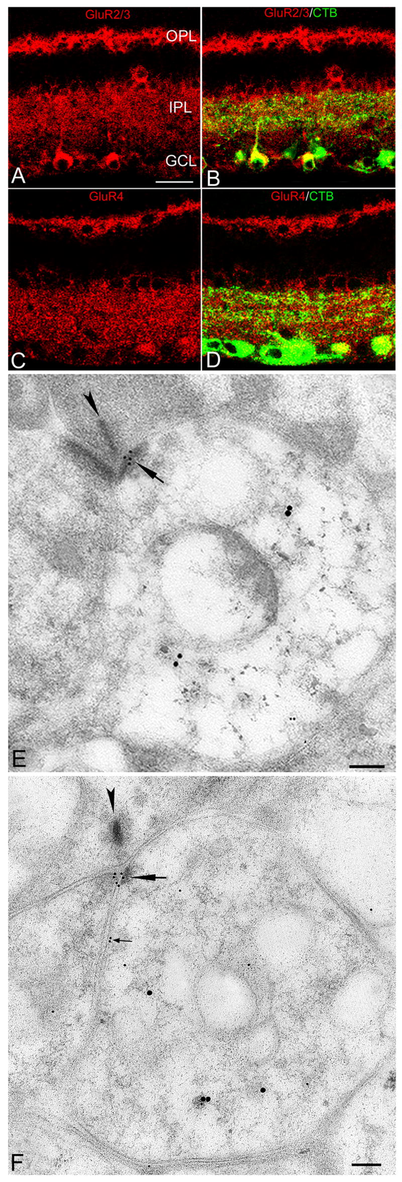
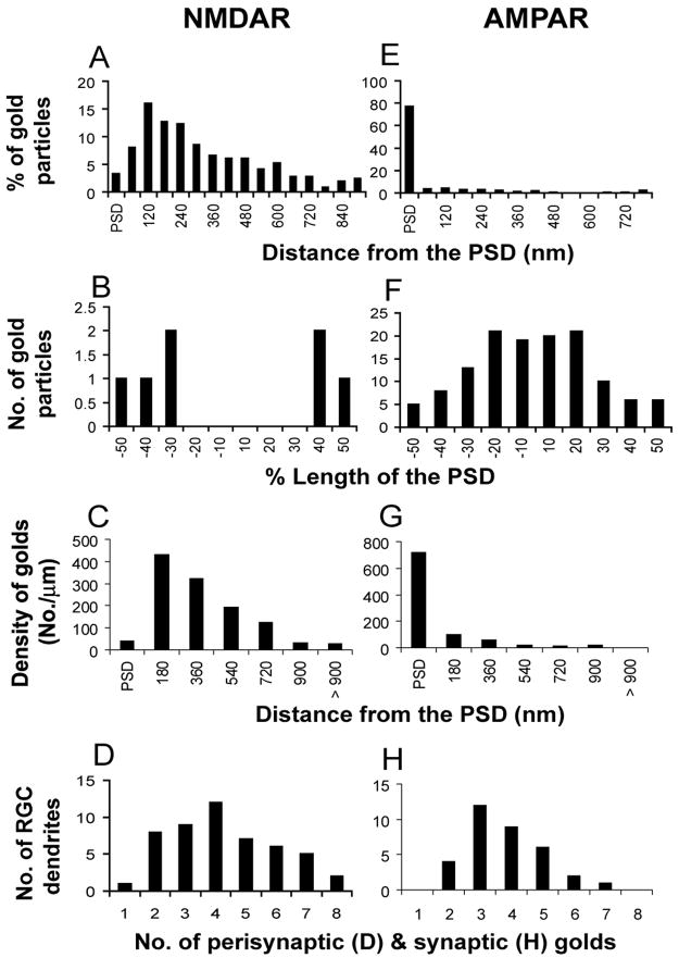
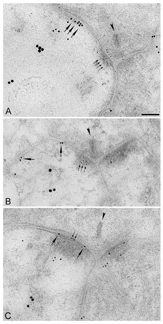
At most excitatory synapses, AMPA and NMDA receptors (AMPARs and NMDARs) occupy the postsynaptic density (PSD) and contribute to miniature excitatory postsynaptic currents (mEPSCs) elicited by single transmitter quanta. Juxtaposition of AMPARs and NMDARs may be crucial for certain types of synaptic plasticity, although extrasynaptic NMDARs may also contribute. AMPARs and NMDARs also contribute to evoked EPSCs in retinal ganglion cells (RGCs), but mEPSCs are mediated solely by AMPARs. Previous work indicates that an NMDAR component emerges in mEPSCs when glutamate uptake is reduced, suggesting that NMDARs are located near the release site but perhaps not directly beneath in the PSD. Consistent with this idea, NMDARs on RGCs encounter a lower glutamate concentration during synaptic transmission than do AMPARs. To understand better the roles of NMDARs in RGC function, we used immunohistochemical and electron microscopic techniques to determine the precise subsynaptic localization of NMDARs in RGC dendrites. RGC dendrites were labeled retrogradely with cholera toxin B subunit (CTB) injected into the superior colliculus (SC) and identified using postembedding immunogold methods. Colabeling with antibodies directed toward AMPARs and/or NMDARs, we found that nearly all AMPARs are located within the PSD, while most NMDARs are located perisynaptically, 100-300 nm from the PSD. This morphological evidence for exclusively perisynaptic NMDARs localizations suggests a distinct role for NMDARs in RGC function.


Figures

Figure 1
Figure 1. Preadsorption with antigen blocks primary antibody immunoreactivity
A: Images of retinal vertical sections showing anti-NR1 labeling pattern in control (A1) and following preadsorption of the antibody with the peptide antigen (A2). Panel A3 shows a representative EM section incubated with preadsorbed primary antibody. B: As in A, but with the anti-NR2B antibody. C: As in A, but with the anti-GluR2/3 antibody. D: As in A, but with the anti-GluR4 antibody. In EM images, presynaptic ribbons are indicated by arrowheads. Scale bars: 20 μm and 0.1 μm for light and electron micrographs, respectively.
Figure 2
Figure 2. RGCs retrogradely labeled with CTB
A–C, Immunofluorescence images of flat-mounted retinas showing CTB-labeled RGCs in the nerve fiber layer (NFL), ganglion cell layer (GCL), and inner plexiform layer (IPL), as well as a small number of displaced RGCs in the inner nuclear layer (INL). D, Immunofluorescence image of transverse retinal sections showing CTB labeling at lower magnification. E, Superimposed immunofluorescence and differential interference contrast images showing CTB labeling in the IPL. Arrow indicates a displaced RGC. F, Co-labeling of CTB and PKC, a marker for rod bipolar cells, indicates that RGC dendrites ramify in the outer and middle thirds of the IPL, while rod bipolar cell terminals occupy the inner third. G, Electron micrograph showing that immunogold particles labeling CTB were excluded from the nucleus (N) of RGCs. H, A CTB-positive RGC dendrite in sublamina a. I and J, CTB-positive postsynaptic processes in sublamina a (I) and b (J). Presynaptic ribbons indicated by arrowheads. Scale bars: 50 μm (A–D), 20 μm (E, F) and 0.1 μm (G–J).
Figure 3
Figure 3. NMDAR immunoreactivity in the rat retina
A and C, NR1 and NR2B immunoreactivity in the IPL, OPL, as well somata in the GCL and INL. B and D, NR1- and NR2B-positive puncta colocalized with CTB-labeled RGC dendrites and somata (yellow). E and F, Electron micrographs showing double immunogold labeling of NMDARs (small particles) and CTB (large particles). At a cone dyad in the OFF sublamina, a cluster of small gold particles (arrow) in the extrasynaptic plasma membrane of RGC dendrites (E); at a dyad in the ON sublamina, two ribbons (arrowheads) each making a dyad with the same CTB-labeled RGC dendrite in which small gold particles (arrow) were on the extrasynaptic plasma membrane (F). Scale bar: 20 μm (A–D), 0.1 μm (E & F).
Figure 4
Figure 4. AMPAR immunoreactivity in the rat retina
A and C, GluR2/3 and GluR4 immunoreactivity in the IPL, OPL, and in somata within the GCL and INL. B and D, GluR2/3 - and GluR4 -positive puncta colocalized with CTB-labeled RGC dendrites and somata (yellow). E and F, Electron micrographs showing double immunogold labeling of AMPARs (small golds) and CTB (large golds) in the OFF and ON sublamina, respectively; small gold particles are clustered (large arrows) in the PSD of RGC processes. F, A rare example of an AMPA-positive RGC process expressing AMPARs perisynaptically (small arrows). Scale bar: 20 μm (A–D), 0.1 μm (E and F).
Figure 5
Figure 5. Quantitative comparison of NMDARs and AMPARs localization in RGC dendrites
A and E, Histograms show the tangential distribution of immunogolds for NMDARs (n=51, A) and AMPARs (n=48, E) within and outside the PSD. The perisynaptic region was divided into 60 nm bins. B and F, Histograms show the tangential distribution of immunogold particles labeling NMDARs (n=4 synapses, B) and AMPARs (n=48 synapses, F) within the PSD. C and G, Histograms show the density of immunogold particles labeling NMDARs (n=51, C) and AMPARs (n=48, G). The perisynaptic region was divided into 180 nm bins. D and H, Histograms showing the number of gold particles detected at NMDAR- and AMPAR-immunopositive synapses, respectively.
Figure 6
Figure 6. Simultaneous labeling of NMDARs, AMPARs and RGCs
Electron micrographs showing triple immunogold labeling of AMPARs (5 nm gold), NMDARs (10 nm gold) and CTB (18 nm gold). A and B, AMPAR gold particles (small arrows) were located in the PSD while NMDAR gold particles (large arrows) were perisynaptic on individual CTB-positive RGC dendrites in the ON and OFF sublamina, respectively. (C) A rare example of NMDARs and AMPARs co-localized within the PSD. Scale bar: 0.1 μm (A–C).

References

    1. Amthor FR, Takahashi ES, Oyster CW. Morphologies of rabbit retinal ganglion cells with complex receptive fields. J Comp Neurol. 1989;280:97–121. - PubMed
    1. Baude A, Nusser Z, Molnar E, McIlhinney RA, Somogyi P. High-resolution immunogold localization of AMPA type glutamate receptor subunits at synaptic and non-synaptic sites in rat hippocampus. Neuroscience. 1995;69:1031–1055. - PubMed
    1. Baude A, Nusser Z, Roberts JD, Mulvihill E, McIlhinney RA, Somogyi P. The metabotropic glutamate receptor (mGluR1 alpha) is concentrated at perisynaptic membrane of neuronal subpopulations as detected by immunogold reaction. Neuron. 1993;11:771–787. - PubMed
    1. Bekkers JM, Stevens CF. NMDA and non-NMDA receptors are co-localized at individual excitatory synapses in cultured rat hippocampus. Nature. 1989;341:230–233. - PubMed
    1. Bernard V, Bolam JP. Subcellular and subsynaptic distribution of the NR1 subunit of the NMDA receptor in the neostriatum and globus pallidus of the rat: co-localization at synapses with the GluR2/3 subunit of the AMPA receptor. Eur J Neurosci. 1998;10:3721–3736. - PubMed

Publication types

MeSH terms

Substances