

The zona pellucida of the koala (Phascolarctos cinereus): its morphogenesis and thickness
- PMID: 16928207
- PMCID: PMC2100332
- DOI: 10.1111/j.1469-7580.2006.00613.x
The zona pellucida of the koala (Phascolarctos cinereus): its morphogenesis and thickness
Abstract
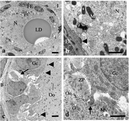
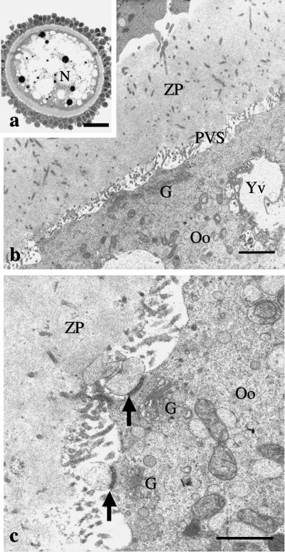
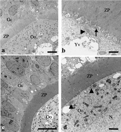
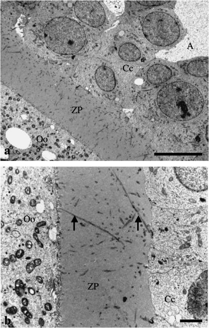
In this study the ultrastructural organization of the koala oocyte and the thickness of the surrounding extracellular coat, the zona pellucida, has been determined to ascertain whether there is coevolution of the morphology of the female gamete with that of the highly divergent male gamete that is found in this marsupial species. Ovaries from several adult koalas were obtained and prepared for transmission electron microscopy. Oocytes in large tertiary follicles were somewhat smaller than those of most other marsupials, although their ultrastructural organization appeared similar and included many yolk vesicles. The zona pellucida surrounding the oocytes in tertiary follicles was approximately 8 microm thick and thus is of similar thickness to that of some eutherian mammals but at least twice as thick as that of most marsupial species so far studied. The results indicate that the koala oocyte is unusually small for a marsupial species whereas the zona pellucida is, by contrast, much thicker. How this relates to sperm-egg interaction at the time of fertilization has yet to be determined.
Figures
Similar articles
-
Ultrastructural characteristics of in vivo and in vitro fertilization in the grey short-tailed opossum, Monodelphis domestica.Anat Rec. 1993 Sep;237(1):21-37. doi: 10.1002/ar.1092370104. Anat Rec. 1993. PMID: 8214640
-
Ultrastructural study of cat zona pellucida during oocyte maturation and fertilization.Anim Reprod Sci. 2008 Nov;108(3-4):425-34. doi: 10.1016/j.anireprosci.2007.09.007. Epub 2007 Oct 2. Anim Reprod Sci. 2008. PMID: 17988809
-
Ultrastructural and histochemical changes in the murine zona pellucida during the final stages of oocyte maturation prior to ovulation.Gamete Res. 1989 Sep;24(1):35-48. doi: 10.1002/mrd.1120240107. Gamete Res. 1989. PMID: 2591851
-
Cytoplasmic maturation of the marsupial oocyte during the periovulatory period.Reprod Fertil Dev. 1996;8(4):509-19. doi: 10.1071/rd9960509. Reprod Fertil Dev. 1996. PMID: 8870076 Review.
-
Zona pellucida characteristics and sperm-binding patterns of in vivo and in vitro produced porcine oocytes inseminated with differently prepared spermatozoa.Theriogenology. 2005 Jan 15;63(2):352-62. doi: 10.1016/j.theriogenology.2004.09.044. Theriogenology. 2005. PMID: 15626404 Review.
Cited by
-
Koala reproduction and the implications for human research.F S Rep. 2025 Apr 15;6(Suppl 1):13-18. doi: 10.1016/j.xfre.2024.12.006. eCollection 2025 Apr. F S Rep. 2025. PMID: 40487325 Free PMC article.
References
-
- Bedford JM. Fertilization. In: Austin CR, Short RV, editors. Reproduction in Mammals, Germ Cells and Fertilization. 2. Vol. 1. Cambridge: Cambridge University Press; 1982. pp. 128–163.
-
- Bedford JM. The coevolution of mammalian gametes. In: Dunbar BS, O'Rand MG, editors. A Comparative Overview of Mammalian Fertilization. New York: Plenum Press; 1991. pp. 3–35.
-
- Bedford JM. What marsupial gametes disclose about gamete function in eutherian mammals. Reprod Fertil Dev. 1996;8:569–580. - PubMed
-
- Bedford JM. Mammalian fertilization misread? Sperm penetration of the eutherian zona pellucida is unlikely to be a lytic event. Biol Reprod. 1998;59:1275–1287. - PubMed
-
- Bedford JM. Enigmas of mammalian gamete form and function. Biol Rev. 2004;79:429–460. - PubMed
Publication types
MeSH terms
LinkOut - more resources
Full Text Sources
