. 2006 Sep;209(3):401-13.
doi: 10.1111/j.1469-7580.2006.00608.x.

Use of a ROSA26:GFP transgenic line for long-term Xenopus fate-mapping studies

Affiliations

Use of a ROSA26:GFP transgenic line for long-term Xenopus fate-mapping studies

Joshua B Gross et al. J Anat. 2006 Sep.

Abstract

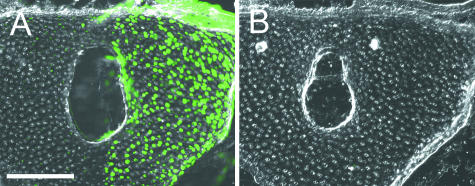
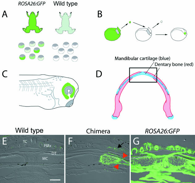
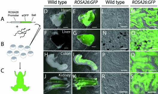
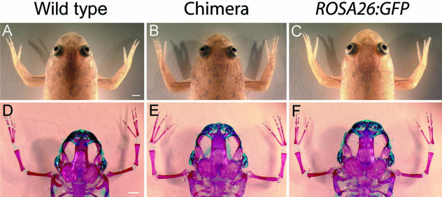
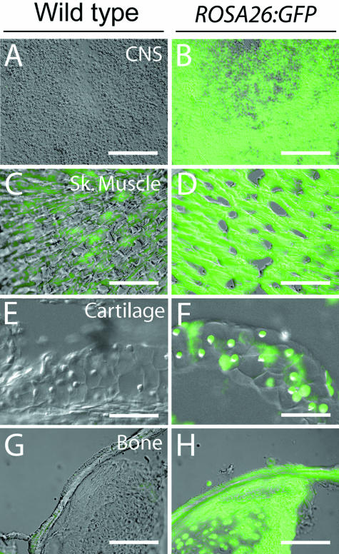
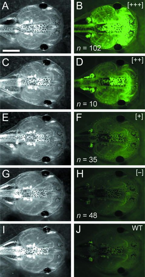
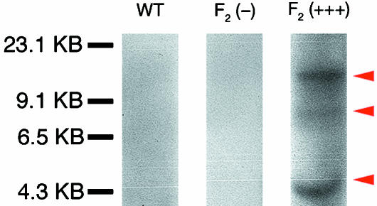
Widespread and persistent marker expression is a prerequisite for many transgenic applications, including chimeric transplantation studies. Although existing transgenic tools for the clawed frog, Xenopus laevis, offer a number of promoters that drive widespread expression during embryonic stages, obtaining transgene expression through metamorphosis and into differentiated adult tissues has been difficult to achieve with this species. Here we report the application of the murine ROSA26 promoter in Xenopus. GFP is expressed in every transgenic tissue and cell type examined at post-metamorphic stages. Furthermore, transgenic ROSA26:GFP frogs develop normally, with no apparent differences in growth or morphology relative to wild-type frogs. ROSA26 transgenes may be used as a reliable marker for embryonic fate-mapping of adult structures in Xenopus laevis. Utility of this transgenic line is illustrated by its use in a chimeric grafting study that demonstrates the derivation of the adult bony jaw from embryonic cranial neural crest.


Figures

Fig 1
Fig 1
Use of a polyclonal anti-GFP antibody significantly amplifies GFP signal in cryosections of chimeric Xenopus. (A,B) Serial cross-sections through the rostral cartilage of an adult frog (NF stage 66, +2 months). Tissue processed using anti-GFP antibody (A; green dots) demonstrates robust staining for GFP. There is no detectable GFP expression in an adjacent section from the same individual (B; separated by ∼60 µm), which was processed without the antibody. In both sections, the right side received a grafted explant of cranial neural crest from a ROSA26:GFP transgenic donor; the left (ungrafted) side of the wild-type host provided an internal control. Scale bar, 200 µm.
Fig 5
Fig 5
Embryonic derivation of the adult lower jaw. We grafted premigratory cranial neural crest (CNC) from transgenic ROSA26:GFP donor embryos to wild-type host embryos at neurula stage, utilizing published fate maps (Sadaghiani & Thiébaud, 1987). In all experiments, eggs from both the ROSA26:GFP founder female and a wild-type female were fertilized in vitro with sperm from a wild-type male (A). ROSA26:GFP zygotes demonstrated variable intensity of GFP fluorescence; such variability is typical in animals with multiple transgene integration sites. Only the brightest, healthiest looking embryos were used as CNC explant donors (B). A tissue explant was removed from a transgenic ROSA26:GFP donor and replaced into a wild-type host. The explant was allowed to heal in place and was assayed the next morning to ensure proper development and migration of the CNC within the mandibular stream (C). Embryonic derivation of the adult mandibular cartilage and dentary bone in the lower jaw was assessed in chimeras that received a labelled graft of the mandibular stream of CNC. The jaw was sectioned in frontal plane (boxed region, D) and assessed for the presence of fluorescent label. GFP staining is absent in a wild-type specimen (E), whereas the entire jaw is labelled strongly in a ROSA26:GFP adult (G). Only the right (operated) side expresses GFP in a chimera that received the labelled graft (F). The left (control) side lacks the label, indicating that graft-derived cells did not cross to the contralateral side of the embryo or adult during development. Dense staining is evident within the extracellular matrix of the dentary bone (red arrowheads). Additional staining is present in the premaxillary bone of the upper jaw as well as portions of several tooth buds (black arrow). Abbreviations: DT, dentary bone; MC, mandibular cartilage; PMx, premaxillary bone; TC, tooth cusps. Scale bar (E–G), 250 µm.
Fig 2
Fig 2
Schematic depiction of REMI method of ROSA26:GFP transgenesis and anti-GFP staining of transgenic tissues. (A) A plasmid containing the ROSA26:GFP cassette is incubated with de-membranated wild-type sperm nuclei in a solution containing restriction enzyme (SalI). Resultant double-stranded breaks in the sperm nuclei genome allow for the introduction of the linearized plasmid. (B) Single nuclei are injected into unfertilized wild-type oocytes. (C) Animals carrying successful integration events and showing widespread GFP fluorescence are reared through metamorphosis. Offspring of a single founder female showing strong fluorescence were used as labelled, cranial neural crest (CNC) explant donors in all experiments. Several tissues were assayed for anti-GFP staining in whole-mounts and serial sections (D–S). In all tissues examined, the ROSA26 promoter drives GFP expression well into adulthood. Two wild-type organs processed for whole-mount immunohistochemistry, heart (D) and kidney (J), showed minor background staining that was much lower than the signal detected in comparable tissues from animals expressing the ROSA26:GFP transgene (E and K, respectively). The background staining was not evident in sections (L, R) when compared with ROSA26:GFP tissue (M, S). Scale bars: D–K, 1 mm; L–S, 100 µm.
Fig 4
Fig 4
External cranial morphology and skeletal development are indistinguishable among wild-type frogs (A,D), ROSA26:GFP transgenic frogs (C,F) and chimeric frogs (B,E). All animals were reared to at least 6 months of age; skeletal anatomy was assessed in cleared-and-stained whole-mounts (D–F). The three groups were indistinguishable from one another in terms of normal development and growth. The minor differences in cranial size among the three cleared-and-stained specimens depicted here are common even among populations of normal tadpoles reared under identical conditions but in different tanks. Scale bar, 2 mm.
Fig 3
Fig 3
Antibody staining of adult cranial tissues. Frontal sections through the prosencephalon (A,B), pterygoideus muscle (C,D), rostral cartilage (E,F) and (ossified) pars articularis of the quadrate bone were immunostained to determine if the transgenic GFP label persists in these tissues. In all comparisons, anti-GFP staining in the ROSA26:GFP tissues (B,D,F,H) is clearly distinguishable when compared with wild-type tissues (A,C,E,G). Wild-type pterygoideus muscle showed minor background fluorescence (C) that is clearly distinguishable from the much more intense positive GFP staining of ROSA26:GFP muscle (D). CNS, central nervous system; Sk. muscle, skeletal muscle. Scale bar, 100 µm.
Fig 6
Fig 6
Variability of endogenous GFP fluorescence among live F2 individuals. Eggs from a single ROSA26 transgenic female (F1) were fertilized in vitro using wild-type sperm. Qualitative were recorded among the resulting F2 progeny viewed under identical conditions (constant time and intensity of exposure to UV illumination). These individuals were assigned among four groups according to qualitative measures of GFP fluorescence: brightest (B; +++), intermediate (D; ++), low (F; +) and undetectable (H; −). The last group is indistinguishable from wild-type individuals (J). There was no apparent difference in morphology among F2 individuals, nor between transgenic and wild-type individuals. The 3 : 1 ratio of GFP-expressing (B,D,F; n = 147) to non-GFP-expressing individuals (H; n = 48) suggests that the F1 female assayed in this experiment probably carries multiple copies of the ROSA26:GFP transgene. Scale bar, 1 mm.
Fig 7
Fig 7
Southern blot analysis of members of the F2 generation of transgenic ROSA26 individuals. Genomic DNA from a wild-type individual (WT), an F2 individual expressing fluorescence at levels indistinguishable from wild-type (−), and an F2 individual expressing the brightest level of GFP fluorescence (+++) were analysed for the presence of transgene integration using Southern blot analysis. Both the wild-type individual and the transgenic F2-individual are shown not to carry the transgene. The F2 (+++) individual, however, carries multiple copies of the transgene (arrowheads). At present, it is not clear if fluorescence is correlated with the site of integration, number of copies at each integration site, or a combination of the two.

Similar articles

Cited by

References

    1. Allen BG, Weeks DL. Transgenic Xenopus laevis embryos can be generated using phiC31 integrase. Nat Meth. 2005;2:975–979. - PMC - PubMed
    1. Balaban E, Teillet M-A, Le Douarin N. Application of the quail-chick chimera system to the study of brain development and behavior. Science. 1988;241:1339–1342. - PubMed
    1. Billington N, Knight AW. Seeing the wood through the trees: a review of techniques for distinguishing green fluorescent protein from endogenous autofluorescence. Anal Biochem. 2001;291:175–197. - PubMed
    1. Chai Y, Jiang X, Ito Y, et al. Fate of the mammalian cranial neural crest during tooth and mandibular morphogenesis. Development. 2000;127:1671–1679. - PubMed
    1. Chalfie M, Tu Y, Euskirchen G, Ward WW, Prasher DC. Green fluorescent protein as a marker for gene expression. Science. 1994;263:802–805. - PubMed

Publication types

LinkOut - more resources