. 2006 Nov;80(21):10900-6.
doi: 10.1128/JVI.00385-06. Epub 2006 Aug 23.

Construction of a severe acute respiratory syndrome coronavirus infectious cDNA clone and a replicon to study coronavirus RNA synthesis

Affiliations

Construction of a severe acute respiratory syndrome coronavirus infectious cDNA clone and a replicon to study coronavirus RNA synthesis

Fernando Almazán et al. J Virol. 2006 Nov.

Abstract

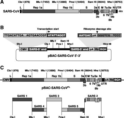
The engineering of a full-length infectious cDNA clone and a functional replicon of the severe acute respiratory syndrome coronavirus (SARS-CoV) Urbani strain as bacterial artificial chromosomes (BACs) is described in this study. In this system, the viral RNA was expressed in the cell nucleus under the control of the cytomegalovirus promoter and further amplified in the cytoplasm by the viral replicase. Both the infectious clone and the replicon were fully stable in Escherichia coli. Using the SARS-CoV replicon, we have shown that the recently described RNA-processing enzymes exoribonuclease, endoribonuclease, and 2'-O-ribose methyltransferase were essential for efficient coronavirus RNA synthesis. The SARS reverse genetic system developed as a BAC constitutes a useful tool for the study of fundamental viral processes and also for developing genetically defined vaccines.


Figures

FIG. 1.
FIG. 1.
Strategy to assemble a SARS-CoV infectious cDNA clone as a BAC. (A) Genetic structure of the SARS-CoV Urbani strain genome. Relevant restriction sites used for the assembly of the full-length cDNA clone are indicated. Numbers in parentheses indicate the genomic positions of the first nucleotide of the restriction endonuclease recognition sequence. Letters and numbers indicate the viral genes. L, leader sequence; UTR, untranslated region; An, poly(A) tail. (B) Construction of pBAC-SARS-CoV 5′-3′. After the selection of appropriate restriction sites, the intermediate plasmid pBAC-SARS-CoV 5′-3′ was constructed as the backbone for assembling the infectious cDNA clone. This plasmid includes the first 681 nt of the genome under the control of the CMV promoter, a multiple-cloning site containing the restriction sites selected for the final assembly of the infectious clone, and the last 975 nt of the genome, followed by a synthetic poly(A) tail (pA), the hepatitis delta virus ribozyme (Rz), and the bovine growth hormone termination and polyadenylation sequences (BGH). All these elements were precisely joined by overlapping PCR. The CMV promoter transcription start and the ribozyme cleavage site are shown. (C) Schematic diagram showing the five-step cloning strategy used for the assembly of the SARS-CoV full-length cDNA clone. The five overlapping cDNA fragments, named SARS 1 to SARS 5, were sequentially cloned into the plasmid pBAC-SARS-CoV 5′-3′ to generate the plasmid pBAC-SARS-CoVFL. Relevant restriction sites are indicated. The labels are as described for panel A.
FIG. 2.
FIG. 2.
Recovery of infectious rSARS-CoV from the full-length cDNA clone. (A) Virus rescue. BHK cells were mock transfected or transfected with either the full-length cDNA clone (pBAC-SARSFL) or a nonreplicative cDNA clone (pBAC-NR) with a deletion in the replicase gene. Cells were removed with trypsin at 6 hpt and plated over a confluent monolayer of VeroE6 cells, and at the indicated times posttransfection, virus titers were determined by plaque assay on VeroE6 cells. Error bars represent standard deviations of the means from three experiments. (B) VeroE6 cells were mock infected or infected with rSARS-CoV and subsequently analyzed for the induction of cytopathic effect (CPE) by light microscopy. Viral protein expression was analyzed by indirect immunofluorescence (IFA) with a human anti-SARS-CoV polyclonal serum, kindly provided by A. Xu (Sun Yat-sen University, Guangzhou, People's Republic of China), followed by fluorescein isothiocyanate-labeled goat anti-human antibody.
FIG. 3.
FIG. 3.
Assembly and functional analysis of a SARS-CoV-derived replicon. (A) Strategy for the construction of the SARS-CoV replicon. A five-step cloning strategy in which the overlapping cDNA fragments, named SARS 1 to SARS 5, were sequentially cloned into the plasmid pBAC-SARS-CoV 5′-3′ to generate plasmid pBAC-SARS-CoV-REP was used. The genetic structure of the replicon and the positions of relevant restriction sites are illustrated. The sequences containing the multiple-cloning site (PacI, AscI, and BamHI) downstream of the replicase gene, and the N gene transcription-regulating sequence (white box) and core sequence (black box) are indicated at the top. Abbreviations are as described in the legend to Fig. 1. (B) Functional analysis of SARS-CoV replicon by RT-PCR. Human 293T and BHK cells were mock transfected or transfected with the replicon (REP) or a nonreplicative construct (NR) by using Lipofectamine 2000 (Invitrogen). Total RNA was isolated at 24 hpt and analyzed by RT-PCR with specific oligonucleotides to detect N gene mRNA. Duplicate RT-PCR products amplified in parallel were resolved by electrophoresis in 1% agarose gels. MW, molecular weight markers. (C) Stability of SARS-CoV replicon in E. coli cells. Plasmid pBAC-SARS-CoV-REP extracted from E. coli DH10B cells that were grown for the indicated number of generations was transfected in 293T cells and the replicon activity analyzed by RT-PCR as described above. Duplicate RT-PCR products amplified in parallel were resolved by electrophoresis in 1% agarose gels. MW, molecular weight markers.
FIG. 4.
FIG. 4.
Construction and functional analysis of a set of SARS-CoV replicons defective in ExoN, NendoU, and 2′-O-MT activities. (A) Schematic diagram showing the genetic structures of the deletion mutant replicons. Relevant restriction sites and the nucleotide positions flanking the deletions from the replicons are indicated. Stop codons are indicated by asterisks. The conserved domains described by Snijder and colleagues (25) are illustrated by the darker bars. Arrowheads indicate the predicted main proteinase (3CL) cleavage sites. Abbreviations above the top bar are as described in the legend to Fig. 1. (B) Functional analysis of mutant replicons by RT-PCR. Human 293T and BHK cells were mock transfected (M) or transfected with the wild-type replicon (REP), the single-deletion mutants (ΔExoN, ΔNendoU, and Δ2′-O-MT), or the deletion mutant lacking the three conserved domains (Δ3). Replicon activity was analyzed by RT-PCR with specific oligonucleotides to detect N gene mRNA. Duplicate RT-PCR products amplified in parallel were resolved by electrophoresis in 1% agarose gels. MW, molecular weight markers. (C) Schematic diagram showing the positions of the point mutations introduced into the ExoN (D6145A), NendoU (H6678A or S6744A), and 2′-O-MT (D6905A) domains. Amino acid numbers refer to their positions in the pp1ab polyprotein. (D) Functional analysis of mutant replicons by real-time RT-PCR. The amount of N gene mRNA, expressed as relative units, was determined by real-time RT-PCR in RNA samples isolated at 24 hpt from BHK cells transfected with the wild-type replicon (REP), the deletion mutant lacking the three conserved domains (Δ3), the single-deletion mutants (ΔExoN, ΔNendoU, and Δ2′-O-MT), or the point mutants (D6145A, H6678A, S6744A, and D6905A mutants). Mean values from three experiments are represented, with standard deviations shown as error bars.

Similar articles

Cited by

References

    1. Adler, H., M. Messerle, and U. H. Koszinowski. 2003. Cloning of herpesviral genomes as bacterial artificial chromosomes. Rev. Med. Virol. 13:111-121. - PubMed
    1. Almazán, F., C. Galán, and L. Enjuanes. 2004. The nucleoprotein is required for efficient coronavirus genome replication. J. Virol. 78:12683-12688. - PMC - PubMed
    1. Almazán, F., J. M. González, Z. Pénzes, A. Izeta, E. Calvo, J. Plana-Durán, and L. Enjuanes. 2000. Engineering the largest RNA virus genome as an infectious bacterial artificial chromosome. Proc. Natl. Acad. Sci. USA 97:5516-5521. - PMC - PubMed
    1. Anand, K., J. Ziebuhr, P. Wadhwani, J. R. Mesters, and R. Hilgenfeld. 2003. Coronavirus main proteinase (3CLpro) structure: basis for design of anti-SARS drugs. Science 300:1763-1767. - PubMed
    1. Brian, D. A., and R. S. Baric. 2005. Coronavirus genome structure and replication. Curr. Top. Microbiol. Immunol. 287:1-30. - PMC - PubMed

Publication types

Associated data

LinkOut - more resources