. 2006 Nov;80(21):10428-35.
doi: 10.1128/JVI.01362-06. Epub 2006 Aug 23.

Generation and analysis of infectious virus-like particles of uukuniemi virus (bunyaviridae): a useful system for studying bunyaviral packaging and budding

Affiliations

Generation and analysis of infectious virus-like particles of uukuniemi virus (bunyaviridae): a useful system for studying bunyaviral packaging and budding

Anna K Overby et al. J Virol. 2006 Nov.

Abstract

In the present report we describe an infectious virus-like particle (VLP) system for the Uukuniemi (UUK) virus, a member of the Bunyaviridae family. It utilizes our recently developed reverse genetic system based on the RNA polymerase I minigenome system for UUK virus used to study replication, encapsidation, and transcription by monitoring reporter gene expression. Here, we have added the glycoprotein precursor expression plasmid together with the minigenome, nucleoprotein, and polymerase to generate VLPs, which incorporate the minigenome and are released into the supernatant. The particles are able to infect new cells, and reporter gene expression can be monitored if the trans-acting viral proteins (RNA polymerase and nucleoprotein) are also expressed in these cells. No minigenome transfer occurred in the absence of glycoproteins, demonstrating that the glycoproteins are absolutely required for the generation of infectious particles. Moreover, expression of glycoproteins alone was sufficient to produce and release VLPs. We show that the ribonucleoproteins (RNPs) are incorporated into VLPs but are not required for the generation of particles. Morphological analysis of the particles by electron microscopy revealed that VLPs, either with or without minigenomes, display a surface morphology indistinguishable from that of the authentic UUK virus and that they bud into Golgi vesicles in the same way as UUK virus does. This infectious VLP system will be very useful for studying the bunyaviral structural components required for budding and packaging of RNPs and receptor binding and may also be useful for the development of new vaccines for the human pathogens from this family.


Figures
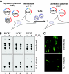
FIG. 1.
FIG. 1.
VLP transfer of UUK minigenomes is dependent on UUK glycoproteins. A. Schematic representation of the experimental setup. BHK-21 cells were transfected with minigenome (M-CAT, L-CAT, or S-CAT) together with the individual expression plasmids pUUK-GN/GC, pUUK-L, and pUUK-N. Supernatant containing VLPs was harvested and inoculated into BHK-21 cells, which were pretransfected with pUUK-L and pUUK-N. Cells were analyzed for reporter gene expression 48 h after VLP infection. Open reading frames are indicated in red, CMV and Pol1 promoters in blue, and NCRs of the M, L, and S in green. B. Comparison of the three different UUK virus-derived minigenomes M-CAT (lanes 1 and 2), L-CAT (lanes 3 and 4), and S-CAT (lanes 5 and 6) with respect to VLP transfer and reporter gene expression (negative controls without pUUK-GN/GC [lanes 1, 3, and 5]). The cell lysates in the CAT reaction were compensated with regard to promoter strength (1x M-CAT, 2x L-CAT, and 4x S-CAT) in both transfected and VLP-infected cells. Transfected cells (top panel) and VLP-infected cells (bottom panel) are shown. Data shown are representative of three independent experiments. C. Transfer of GFP-containing minigenome (M-GFP) by UUK VLPs. BHK-21 cells were transfected with M-GFP and the L, N, and GN/GC expression plasmids (top panel) or infected with GFP-containing VLPs (bottom panel).
FIG. 2.
FIG. 2.
Optimization of VLP formation. A. Different amounts of pUUK-GN/GC (0, 0.5, 1.0, 2.5, and 5.0 μg) were transfected together with the other plasmids (M-CAT, pUUK-L, and pUUK-N), and at 24 h posttransfection, cell lysates were analyzed for CAT activity (top panel; transfected) and supernatants were used to infect new cells previously transfected with pUUK-L and pUUK-N (bottom panel; VLP infected). VLP-infected cells were analyzed 48 h postinfection. B. Kinetics of VLP formation. Cells were transfected with minigenome and pUUK-L, pUUK-N, and pUUK-GN/GC for the indicated times, analyzed for minigenome activity (top panel), and supernatants containing VLPs were collected. The collected supernatants were passaged to new cells and analyzed for CAT activity 48 h after VLP infection (bottom panel). C. Serial passage of VLPs. Cells were transfected with the M-CAT and plasmids expressing L and N proteins (top panel, lane 1) or with L, N, and GN/GC proteins (bottom panel, lane 1), and supernatants were passaged three times to new cells 24 h before infection with the VLP supernatant from cells transfected with pUUK-L, pUUK-N, and pUUK-GN/GC. Before CAT activity was analyzed 48 h posttransfection, supernatant was transferred to new cells. P1, P2, and P3 indicate the first, second, and third passages, respectively. Panels A, B, and C show representative CAT pictures from three independent experiments. Background spots from BHK-21 cells are indicated by the black asterisks.
FIG. 3.
FIG. 3.
Inhibition of VLP infection by UUK virus-specific antibodies. Different amounts (100 and 250 μl) of supernatants with VLPs containing the M-CAT minigenome were incubated with 10 or 20 μl of UUK virus immune sera recognizing GN/GC (lanes 5 and 6 and lanes 7 and 8, respectively) or with 20 μl preimmune (pre) serum (lanes 3 and 4) before inoculation onto new cells. The inoculating material was removed 1 h later, and CAT activity was measured 48 h after VLP infection. Untreated supernatants are shown in lanes 1 and 2. The neutralizing effect was seen in three independent experiments.
FIG. 4.
FIG. 4.
Western blot analysis of UUK virus and UUK VLPs released in the supernatant. Supernatants from UUK virus-infected cells (lane 1) and cells transfected with different combinations of UUK plasmids (lanes 2 to 8) were concentrated and analyzed by Western blotting using antibodies specifically recognizing UUK glycoproteins (GN/GC) and nucleoprotein (N).
FIG. 5.
FIG. 5.
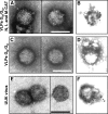
Morphology of the VLPs and UUK virus by transmission electron microscopy. Supernatant from cells transfected with pUUK-GN/GC, pUUK-L, pUUK-N, and M-CAT (A and B) or with only pUUK-GN/GC (C and D) or infected with UUK virus (E and F) were harvested and concentrated through a sucrose cushion. Particle morphology (A, C, and E) was analyzed by negative staining on glutaraldehyde-fixed particles. UUK glycoproteins were visualized by immunogold staining (B, D, and F) on unfixed particles using polyclonal antibodies recognizing GN/GC detected with secondary antibodies coupled to 15-nm gold particles. Bars, 100 nm.
FIG. 6.
FIG. 6.
Morphology of VLP-producing BHK-21 cells by thin-section transmission electron microscopy. Cells transfected with pUUK-GN/GC, pUUK-L, pUUK-N, and M-CAT (A and B) or with only pUUK-GN/GC (C and D) or infected with UUK virus (E and F) at 24 h posttransfection or infection were fixed and embedded for TEM. Bars, 100 nm.

References

    1. Ako-Adjei, D., M. C. Johnson, and V. M. Vogt. 2005. The retroviral capsid domain dictates virion size, morphology, and coassembly of Gag into virus-like particles. J. Virol. 79:13463-13472. - PMC - PubMed
    1. Andersson, A. M., L. Melin, A. Bean, and R. F. Pettersson. 1997. A retention signal necessary and sufficient for Golgi localization maps to the cytoplasmic tail of a Bunyaviridae (Uukuniemi virus) membrane glycoprotein. J. Virol. 71:4717-4727. - PMC - PubMed
    1. Barr, J. N., R. M. Elliott, E. F. Dunn, and G. W. Wertz. 2003. Segment-specific terminal sequences of Bunyamwera bunyavirus regulate genome replication. Virology 311:326-338. - PubMed
    1. Barr, J. N., J. W. Rodgers, and G. W. Wertz. 2006. Identification of the Bunyamwera bunyavirus transcription termination signal. J Gen. Virol. 87:189-198. - PubMed
    1. Betenbaugh, M., M. Yu, K. Kuehl, J. White, D. Pennock, K. Spik, and C. Schmaljohn. 1995. Nucleocapsid- and virus-like particles assemble in cells infected with recombinant baculoviruses or vaccinia viruses expressing the M and the S segments of Hantaan virus. Virus Res. 38:111-124. - PubMed

LinkOut - more resources