. 2006 Nov;80(21):10579-90.
doi: 10.1128/JVI.00941-06. Epub 2006 Aug 23.

Viral and cellular determinants of the hepatitis C virus envelope-heparan sulfate interaction

Affiliations

Viral and cellular determinants of the hepatitis C virus envelope-heparan sulfate interaction

Heidi Barth et al. J Virol. 2006 Nov.

Abstract

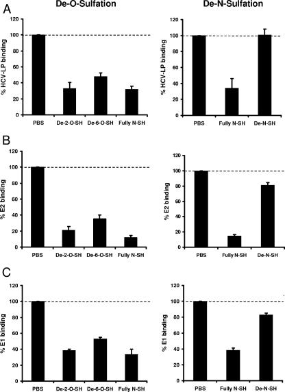
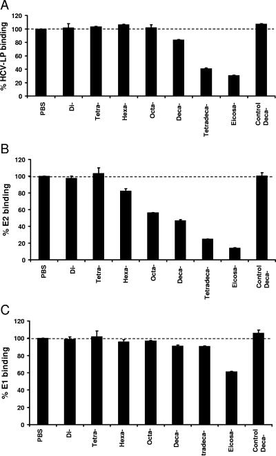
Cellular binding and entry of hepatitis C virus (HCV) are the first steps of viral infection and represent a major target for antiviral antibodies and novel therapeutic strategies. We have recently demonstrated that heparan sulfate (HS) plays a key role in the binding of HCV envelope glycoprotein E2 to target cells (Barth et al., J. Biol. Chem. 278:41003-41012, 2003). In this study, we characterized the HCV-HS interaction and analyzed its inhibition by antiviral host immune responses. Using recombinant envelope glycoproteins, virus-like particles, and HCV pseudoparticles as model systems for the early steps of viral infection, we mapped viral and cellular determinants of HCV-HS interaction. HCV-HS binding required a specific HS structure that included N-sulfo groups and a minimum of 10 to 14 saccharide subunits. HCV envelope binding to HS was mediated by four viral epitopes overlapping the E2 hypervariable region 1 and E2-CD81 binding domains. In functional studies using HCV pseudoparticles, we demonstrate that HCV binding and entry are specifically inhibited by highly sulfated HS. Finally, HCV-HS binding was markedly inhibited by antiviral antibodies derived from HCV-infected individuals. In conclusion, our results demonstrate that binding of the viral envelope to a specific HS configuration represents an important step for the initiation of viral infection and is a target of antiviral host immune responses in vivo. Mapping of viral and cellular determinants of HCV-HS interaction sets the stage for the development of novel HS-based antiviral strategies targeting viral attachment and entry.


Figures

FIG. 1.
FIG. 1.
SPR analysis of envelope glycoprotein E1-heparin binding. (A) Heparin-BSA or BSA was covalently immobilized onto the surface of a biosensor chip as described recently (2). Subsequently, different concentrations of recombinant highly purified envelope glycoprotein E1 were injected onto the biosensor surface. The biosensor chip response is indicated on the y axis (measured in RU) as a function of time (x axis) at 25°C. The sensorgram shows the difference in the BSA-heparin coated chip response compared to the BSA-coated control chip response following E1 injection. (B) Side-by-side analysis of E1 and E2 (both at 100 nM).
FIG. 2.
FIG. 2.
Cellular binding of envelope glycoproteins E1 and E2 to human hepatoma cells is HS dependent. (A) Dose-dependent binding of recombinant E1 and E2 to HepG2 and Huh-7 cells. Cells were incubated with increasing concentrations of E1 or E2. Binding of E1 and E2 was analyzed by flow cytometry using a mouse anti-E1 (11B7) or anti-E2 (16A6) MAb, respectively, and PE-conjugated anti-mouse IgG. On the y axis, net mean fluorescence intensity (ΔMFI) values for each protein concentration were calculated by subtracting the MFI of negative controls (cells incubated in PBS without envelope protein and the addition of anti-envelope MAb and PE-conjugated anti-mouse IgG antibodies) from that obtained with cells incubated with envelope proteins at the concentration indicated on the x axis. Data are shown as mean ΔMFI ± standard deviation (SD) of three (E1 and E2 at 50 and 100 μg/ml) or four (all other envelope protein concentrations) experiments. Significant differences in ΔMFIs obtained for E2 versus E1 binding are indicated by asterisks (*, P < 0.05; **, P < 0.01; ***, P < 0.001 [determined by two-tailed t test]). (B) Binding of E1 and E2 to human hepatoma (Huh-7 and HepG2), human nonhepatoma (293T, HeLa, and OKF6), and mouse hepatoma (Hepa 1-6) cell lines. Cells were incubated with recombinant envelope glycoproteins (2.5 μg/ml), and cellular binding of envelope glycoproteins was quantified by flow cytometry in side-by-side experiments as described in panel A. ΔMFI ± SD of a representative experiment performed in triplicate is shown. (C) Flow cytometry histograms of E1 binding to Huh-7 cells (black line, unshaded peak) in the presence of soluble GAGs. Recombinant E1 protein was preincubated with PBS, chondroitin sulfate A (CSA), normally sulfated HS (HS), highly sulfated HS (hsHS), or heparin (each at 10 μg/ml) for 30 min at room temperature. E1-GAG complexes were added to cells, and cellular binding of E1 was quantified by flow cytometry as described for panel A. Background fluorescence (gray-shaded peak) corresponds to cells incubated without envelope protein. (D) Percent cellular binding of E1 protein to HepG2 and Huh-7 in the presence of soluble GAGs relative to binding of E1 without GAGs (100%). Mean ± SD of a representative experiment performed in triplicate is shown.
FIG. 3.
FIG. 3.
Cellular binding of HCV-LPs and recombinant envelope glycoprotein requires N sulfation of cell surface HS. Sucrose gradient-purified HCV-LPs (A) and recombinant E2 (B) and E1 (C) proteins were incubated with chemically modified heparins (10 μg/ml) for 30 min at room temperature. Envelope protein-GAG complexes were added to human hepatoma cells for 1 h at 4°C, and cellular binding of HCV-LPs and recombinant proteins was quantified by flow cytometry as described for Fig. 2A. Data are shown as percent binding of ligands (mean ± standard deviation [SD] of a representative experiment performed in triplicate) relative to binding of ligands in the absence of modified heparins (100%). De-2-O- and de-6-O-sulfated heparin (SH) lacks O-sulfo groups at position 2 or 6; de-N-SH lacks N-sulfo groups.
FIG. 4.
FIG. 4.
Cellular binding of HCV-LPs and recombinant envelope E1 and E2 in the presence of heparin oligosaccharides. HCV-LPs (A), recombinant E2 (B), and E1 protein (C) were preincubated withheparin-derived oligosaccharides ranging in size from di- to eicosasaccharides or a control decasaccharide containing the pentasaccharide antithrombin-III binding site (each at 50 μg/ml) for 30 min at room temperature. Viral protein-oligosaccharide complexes were added to human hepatoma cells, and cellular binding of viral proteins was quantified by flow cytometry using anti-envelope MAb as described for Fig. 2A. Data are shown as percent cellular viral protein binding relative to binding of viral proteins without oligosaccharides (100%).
FIG. 5.
FIG. 5.
Mapping of viral epitopes interacting with HS using an E2-heparin binding assay. (A) Inhibition of E2-heparin binding by monoclonal anti-E2 antibodies. E2 (1 μg/ml) was preincubated with the anti-E2 MAbs or IgG (50 μg/ml) for 1 h at 37°C and then added to ELISA plates coated with heparin (10 μg/ml). Heparin-bound E2 was detected using a polyclonal anti-E2 rabbit serum and colorimetric reaction as described in Materials and Methods. Results are shown as percent inhibition of E2-heparin interaction. Data are shown as mean percent inhibition of E2-heparin binding ± standard deviation (SD) obtained from three independent experiments. (B) Concentration-dependent inhibition of E2-heparin binding by anti-E2 antibodies. Recombinant E2 protein (1 μg/ml) was incubated with anti-E2 monoclonal antibody 2F10 (squares) or 49F3 (diamonds) or with control IgG (open triangles) at various concentrations for 1 h at 37°C. E2-antibody complexes were added to heparin immobilized on plates, and the E2-heparin interaction was analyzed as described above. The OD of the colorimetric reaction is proportional to heparin-bound E2. Results are presented as mean OD ± SD of a representative experiment performed in triplicate.
FIG. 6.
FIG. 6.
Inhibition of envelope glycoprotein binding to immobilized heparin by purified human anti-HCV IgG. (A) Envelope glycoproteins E1 and E2 were incubated with IgG purified from sera from anti-HCV-positive patients (pt1 to pt12) or healthy individuals (control [C]). Envelope glycoprotein-antibody complexes were added to immobilized heparin, and bound envelope glycoproteins were detected by rabbit anti-E1 or anti-E2 polyclonal serum as described in the legend to Fig. 5. Data are shown as mean percent inhibition of E2-heparin binding ± standard deviation (SD) obtained from three independent experiments. To study inhibition of HCVpp entry in the presence of anti-HCV IgG, HCVpp (HCV-J strain) were incubated with purified IgG from HCV-infected patients and human control IgG before the addition to Huh-7 cells. HCVpp entry was analyzed by flow cytometry as described in Materials and Methods. (B) Concentration-dependent inhibition of E2-heparin binding by purified anti-HCV IgG (pt12; solid diamonds) and control IgG (open squares). Analysis of inhibition was performed as described in the legend to Fig. 5. Results are presented as mean OD ± SD of a representative experiment performed in triplicate.
FIG. 7.
FIG. 7.
Inhibition of HCVpp infection of Huh-7 cells by heparin and highly sulfated HS. (A) For HCVpp infection, sucrose gradient-purified HCVpp (HCV-J strain) were preincubated with PBS, heparin, highly sulfated HS (hsHS), normally sulfated HS (HS), or chondroitin sulfate A (CSA) (each at 10 μg/ml) for 30 min at room temperature. HCVpp-GAG complexes were added to Huh-7 cells and incubated for 1.5 h at 4°C. HCVpp entry was determined by GFP reporter gene expression using flow cytometry. Data are shown as mean percent cells positive for GFP relative to infection of HCVpp without GAG (100%). (B) To study whether soluble GAGs interfere with the first step of HCVpp infection, HCVpp binding, HCVpp-GAG complexes were added to Huh-7 cells for 1 h at 4°C and cellular binding of sucrose gradient-purified HCVpp was quantified by anti-E2 MAb (16A6) and flow cytometry. Data are shown as percent binding (mean ± standard deviation [SD] of a representative experiment performed in triplicate) relative to binding of HCVpp without GAG (100%). (C) To study whether soluble GAGs interfere with viral entry mechanisms following viral attachment (temperature-dependent postbinding events), Huh-7 cells were first incubated with HCVpp for 1 h at 4°C. Following binding of HCVpp and washing of cells with PBS to remove unbound HCVpp, soluble GAGs were added to cells (1.5 h at 4°C). Following a shift of incubation temperature to 37°C, HCVpp entry was assessed after 72 h by flow cytometric analysis of GFP reporter gene expression. Data are shown as mean percent cells positive for GFP relative to infection of HCVpp without GAG (100%).

References

    1. Bain, C., A. Fatmi, F. Zoulim, J. P. Zarski, C. Trepo, and G. Inchauspe. 2001. Impaired allostimulatory function of dendritic cells in chronic hepatitis C infection. Gastroenterology 120:512-524. - PubMed
    1. Barth, H., C. Schafer, M. I. Adah, F. Zhang, R. J. Linhardt, H. Toyoda, A. Kinoshita-Toyoda, T. Toida, T. H. Van Kuppevelt, E. Depla, F. Von Weizsacker, H. E. Blum, and T. F. Baumert. 2003. Cellular binding of hepatitis C virus envelope glycoprotein E2 requires cell surface heparan sulfate. J. Biol. Chem. 278:41003-41012. - PubMed
    1. Bartosch, B., J. Dubuisson, and F. L. Cosset. 2003. Infectious hepatitis C virus pseudo-particles containing functional E1-E2 envelope protein complexes. J. Exp. Med. 197:633-642. - PMC - PubMed
    1. Bartosch, B., A. Vitelli, C. Granier, C. Goujon, J. Dubuisson, S. Pascale, E. Scarselli, R. Cortese, A. Nicosia, and F. L. Cosset. 2003. Cell entry of hepatitis C virus requires a set of co-receptors that include the CD81 tetraspanin and the SR-B1 scavenger receptor. J. Biol. Chem. 278:41624-41630. - PubMed
    1. Baumert, T. F., S. Ito, D. T. Wong, and T. J. Liang. 1998. Hepatitis C virus structural proteins assemble into viruslike particles in insect cells. J. Virol. 72:3827-3836. - PMC - PubMed

Publication types

MeSH terms

LinkOut - more resources