. 2006 Aug 23;26(34):8819-28.
doi: 10.1523/JNEUROSCI.2593-06.2006.

The Chk1/Cdc25A pathway as activators of the cell cycle in neuronal death induced by camptothecin

Affiliations

The Chk1/Cdc25A pathway as activators of the cell cycle in neuronal death induced by camptothecin

Yi Zhang et al. J Neurosci. .

Abstract

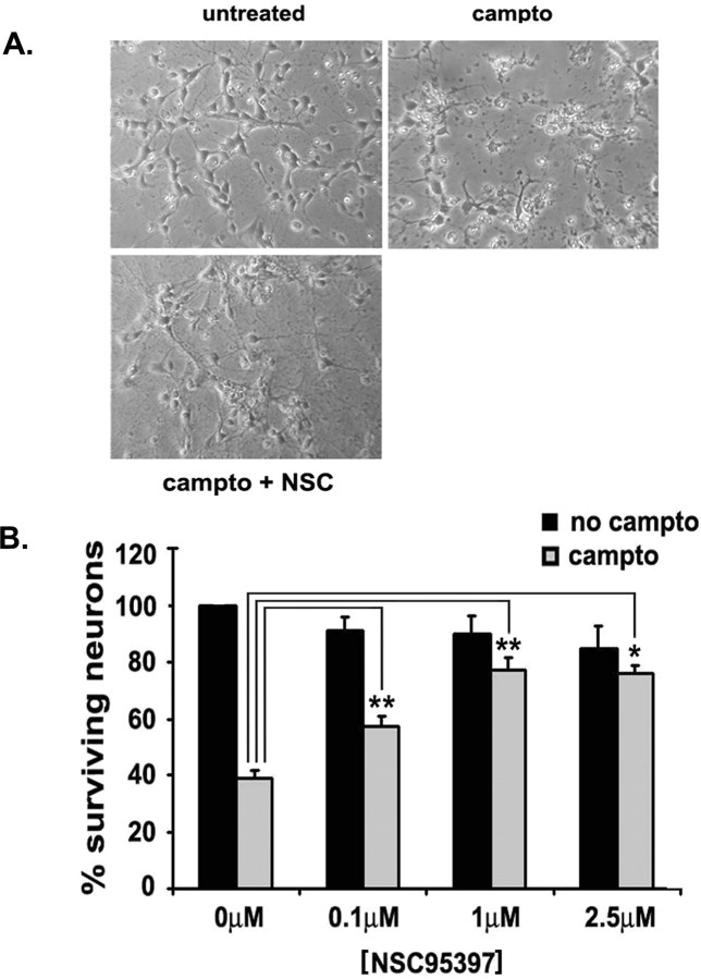
Cell cycle regulators appear to play a paradoxical role in neuronal death. We have shown previously that cyclin-dependent kinases (CDKs), along with their downstream effectors, Rb (retinoblastoma) and E2F/DP1 (E2 promoter binding factor/deleted in polyposis 1), regulate neuronal death evoked by the DNA damaging agent camptothecin. However, the mechanism by which CDKs are activated in this model is unclear. The cell division cycle 25A (Cdc25A) phosphatase is a critical regulator of cell cycle CDKs in proliferating cells. In cortical neurons, we presently show that expression of Cdc25A promotes death even in the absence of DNA damage. Importantly, Cdc25A activity is rapidly increased during DNA damage treatment. Inhibition of Cdc25A blocks death and reduces cyclin D1-associated kinase activity and Rb phosphorylation. This indicates that endogenous Cdc25A activity is important for regulation of cell cycle-mediated neuronal death. We also examined how Cdc25A activity is regulated after DNA damage. Cultured embryonic cortical neurons have a significant basal activity of checkpoint kinase 1 (Chk1), a kinase that regulates cell cycle arrest. During camptothecin treatment of neurons, this activity is rapidly downregulated with a concomitant increase in Cdc25A activity. Importantly, expression of wild-type Chk1, but not kinase-dead Chk1, inhibits the camptothecin-induced increase in Cdc25A activity. In addition, Chk1 expression also promotes survival in the presence of the DNA-damaging agent. Together, our data suggest that a Chk1/Cdc25A activity participates in activation of a cell cycle pathway-mediated death signal in neurons. These data also define how a proliferative signal may be abnormally activated in a postmitotic environment.


Figures

Figure 1.
Figure 1.
Cdc25A phosphatase activity is increased in cortical neurons treated with camptothecin. A, Cdc25A phosphatase was immunoprecipitated from 500 μg of whole-cell lysate of cortical neurons treated for various times with camptothecin (Campto) and subjected to phosphatase assay using pNPP as substrate. Phosphatase activity was assayed by liberation of the pNPP cleavage product quantitated at OD410 by spectrophotometry. Significance comparisons with time 0 control, *p < 0.05. Data represent the mean ± SEM from seven independent experiments. B, A parallel IP/Western blot was performed to demonstrate pull-down of Cdc25A. IgG control refers to a control IP performed with nonspecific IgG control antibody. A cross-reactive IgG band is also visible after Western blot analyses. C, Neurons were fixed and immunostained for Cdc25A, and nuclei were stained with Hoescht. Culture without primary antibody (Ab) incubation was provided as a control.
Figure 2.
Figure 2.
Cdc25A promotes neuronal death in response to DNA damage. Cultured cortical neurons were cotransfected with pEGFP and the indicated constructs. At 24 h after transfection, cells were exposed to camptothecin (campto) (10 μm) for 16–20 h, fixed, and stained with Hoechst 33258 (0.25 μg/ml). A, Photomicrographs of alive or dead cortical neurons under green fluorescence, showing GFP-transfected neurons and Hoechst-stained nuclei. B, Expression of Cdc25A using a plasmid construct induces death of primary cultured cortical neurons treated with camptothecin. Significance comparisons with respective vector untreated or camptothecin-treated controls, **p < 0.01. Data represent the mean ± SEM from three independent experiments.
Figure 3.
Figure 3.
Cdc25A inhibition with NSC95397 protects cortical neurons from camptothecin-induced neuronal death. At 36 h after plating, cortical neurons were treated with camptothecin (10 μm) alone or cotreated with NSC95397. A, Phase micrographs of neuronal cultures not treated, treated with camptothecin (campto) (10 μm) alone, and cotreated with NSC95397 (NSC) (1 μm) for 12 h. B, Survival assay of camptothecin-induced death of cortical neurons cotreated with NSC95397 at the indicated concentrations. Cells were lysed and intact nuclei were counted according to the method outlined in Materials and Methods. Significance comparisons between untreated and camptothecin treatment at each NSC concentration, *p < 0.05, **p < 0.01. Data represent the mean ± SEM from at least three independent experiments.
Figure 4.
Figure 4.
Cdc25A siRNA protects cortical neurons from death evoked by camptothecin. Cortical neurons were transfected with a control siRNA (sicontrol) or two separate Cdc25A siRNA oligonucleotides (siCdc25A1, siCdc25A2) as indicated for 48 h and then treated with camptothecin (campto) for 12 and 16 h also as indicated. Neurons were lysed and numbers of surviving cells were assessed. Data represent means ± SEM of three experiments. Significance comparisons with untreated or camptothecin-treated control siRNA. *p < 0.05, **p < 0.01. Insets at top show Western blot analyses, showing downregulation of Cdc25A in cells treated with siCdc25A1 (A1) or siCdc25A2 (A2) compared with control siRNA (C).
Figure 5.
Figure 5.
Inhibition of Cdc25 phosphatase activity attenuates camptothecin-evoked cyclin D1-associated kinase activity and pRb phosphorylation at Ser795. A, NSC95397 decreases camptothecin-induced cyclin D1-associated kinase activity in cortical neurons. B, Densitometric analysis of cyclin D1-assciated kinase activity. The kinase activity was quantified by measuring the incorporation of 32P onto pRb substrate and subtracting the control background value. Data are the mean ± SEM and are expressed relative to the initial amount of phosphorylated pRb at time 0. C, NSC95397 (1 μm) attenuates the camptothecin-evoked increase in phosphorylation of Rb–Ser795. Western blotting analysis was performed using anti-pRb–Ser795 antibody. Actin is provided as loading control. D, Densitometric analysis of Western immunoblots. Data are the mean ± SEM and are expressed relative to the initial amount of phosphorylated pRb at time 0. E, Cdc25A siRNA (siCdc25A) attenuates the camptothecin (campto)-evoked increase in phosphorylation of Rb–Ser795. Actin is provided as loading control. Similar results were observed in two separate experiments.
Figure 6.
Figure 6.
Cdc25A and Chk1 protein levels do not change significantly in response to DNA damage. Western blot showing relative Cdc25A and Chk1 levels from the whole-cell lysates of cortical neurons after camptothecin (campto) (10 μm) treatment for the indicated times. Cultured cortical neurons were treated with camptothecin (10 μm) for various times 36 h after plating, whole-cell lysates were harvested, and Western blot analysis was performed according to standard procedure. Actin is provided as loading controls.
Figure 7.
Figure 7.
Chk1 kinase activity is rapidly decreased in response to DNA damage. Chk1 activity was calculated by measuring the incorporation of 32P onto GST–Cdc25A substrate and subtracting the control (cont) background value. The kinase activity from three independent experiments were analyzed by densitometry. A, An example of autoradiogram showing the incorporation of 32P into Cdc25A. campto, Camptothecin. B, Autoradiograms were quantified by densitometry. Data represent the mean ± SEM from three independent experiments.
Figure 8.
Figure 8.
Chk1 expression protects neurons from camptothecin-induced death through downregulation of Cdc25A activity. A, The effects of Chk1 on survival of cultured cortical neurons treated with camptothecin (campto). At the time of plating, neuronal cultures were infected with a control virus and both WT-Chk1 and kinase-dead DN-Chk1 viruses (multiplicity of infection, 100) and were then treated with or without camptothecin for 16 h. Significance comparisons with control virus alone, **p < 0.01. B, Cdc25A phosphatase activity with overexpression of WT-Chk1 and DN-Chk1 viruses. The phosphatase activity was normalized to IP control. Significance comparisons with control virus alone untreated or treated with camptothecin for 2 h, *p < 0.05; **p < 0.01.

References

    1. Alam ZI, Jenner A, Daniel SE, Lees AJ, Cairns N, Marsden CD, Jenner P, Halliwell B. Oxidative DNA damage in the parkinsonian brain: an apparent selective increase in 8-hydroxyguanine levels in substantia nigra. J Neurochem. 1997;69:1196–1203. - PubMed
    1. Aleyasin H, Cregan SP, Iyirhiaro G, O’Hare MJ, Callaghan SM, Slack RS, Park DS. Nuclear factor-κB modulates the p53 response in neurons exposed to DNA damage. J Neurosci. 2004;24:2963–2973. - PMC - PubMed
    1. Baratte B, Meijer L, Galaktionov K, Beach D. Screening for antimitotic compounds using the cdc25 tyrosine phosphatase, an activator of the mitosis-inducing p34cdc2/cyclin Bcdc13 protein kinase. Anticancer Res. 1992;12:873–880. - PubMed
    1. Becker EB, Bonni A. Cell cycle regulation of neuronal apoptosis in development and disease. Prog Neurobiol. 2004;72:1–25. - PubMed
    1. Biswas SC, Liu DX, Greene LA. Bim is a direct target of a neuronal E2F-dependent apoptotic pathway. J Neurosci. 2005;25:8349–8358. - PMC - PubMed

Publication types