. 2006 Aug 23;26(34):8866-74.
doi: 10.1523/JNEUROSCI.5191-05.2006.

Axon guidance by diffusible chemoattractants: a gradient of netrin protein in the developing spinal cord

Affiliations

Axon guidance by diffusible chemoattractants: a gradient of netrin protein in the developing spinal cord

Timothy E Kennedy et al. J Neurosci. .

Abstract

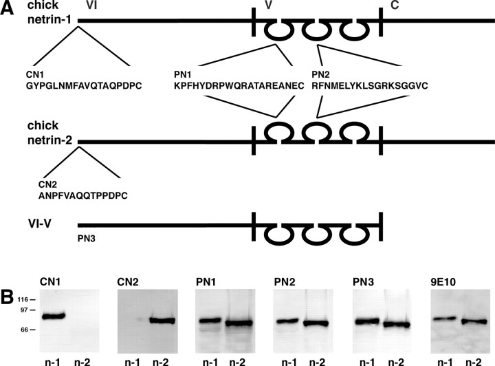
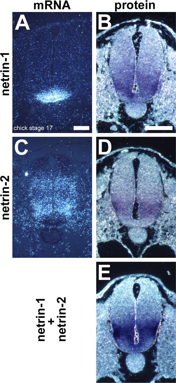
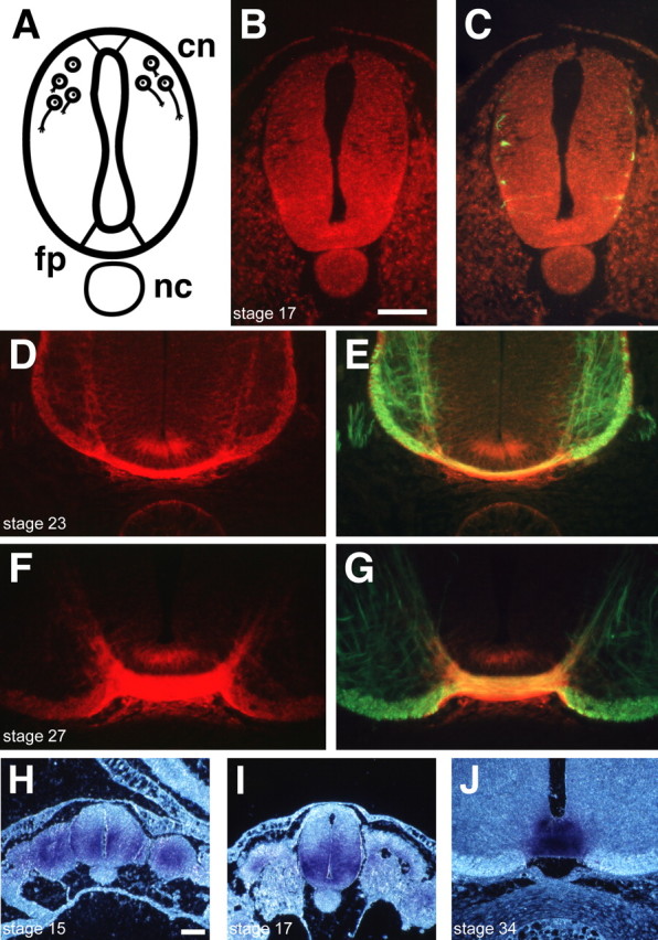
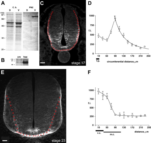
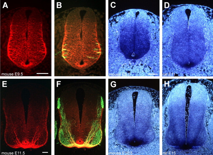
Gradients of diffusible long-range attractant and repellent proteins have been proposed to guide growing axons during nervous system development, but such gradients have never been visualized directly. In the embryonic spinal cord, commissural axons pioneer a circumferential trajectory to the floor plate at the ventral midline directed by secreted proteins of the netrin family. In the embryonic chick spinal cord netrin-1 mRNA is expressed by floor plate cells and netrin-2 mRNA by neural epithelial cells. Antibodies to the two netrins reveal a gradient of netrin protein directly in the path of commissural axons. The netrin-1 gradient itself extends many cell diameters dorsal to the floor plate, the site of netrin-1 expression. A similar distribution of netrin-1 protein has been detected in embryonic rat and mouse spinal cord. The detection of a gradient of netrin-1 protein supports the operation of long-range chemotropic mechanisms in the developing nervous system.


Figures

Figure 1.
Figure 1.
Specificity of netrin antibodies. A, Peptide antigens were synthesized that correspond to sequences derived from the mature N terminus of chick netrin-1 (CN1) and chick netrin 2 (CN2). These sequences are unique to chick netrin-1 and chick netrin-2, respectively. Peptides also were synthesized that correspond to sequences within the domain V EGF-like loops (PN1, PN2) that are conserved in vertebrate netrin-1, netrin-2, and netrin-3 sequences. The PN1 peptide sequence is identical to the corresponding sequence of chick, rat, and mouse netrin-1 and contains a single conserved amino acid substitution in chick netrin-2. PN1 is poorly conserved in mouse netrin-3 and netrin-4, containing six of 20 and 17 of 20 amino acid substitutions, respectively. The PN2 sequence is identical in netrin-1 and netrin-2 in chick, rat, and mouse netrin-1. Two conservative amino acid substitutions are present in mouse netrin-3, and the corresponding sequence in mouse netrin-4 is poorly conserved, containing 10 of 18 amino acid substitutions. Recombinant chicken netrin-1 domain VI–V (PN3) is a 429 amino acid sequence that shares ∼90% amino acid identity with mouse netrin-1, ∼75% amino acid identify with chick netrin-2, ∼57% amino acid identity with mouse netrin-3, and ∼35% amino acid identity with mouse netrin-4. B, Antibody specificity was assessed by using purified recombinant netrin protein and Western blot analysis. In total, 50 ng of purified recombinant full-length myc-tagged chick netrin-1 (n-1) or chick netrin-2 (n-2) protein was loaded per lane. The 9E10 monoclonal antibody against a myc epitope tag confirmed that approximately the same amount of recombinant protein was loaded in each lane. Molecular weight markers, indicated with black bars on the left, correspond to 116, 97, and 66 kDa. Antibodies raised against peptide epitopes (CN1) and (CN2) are specific for chick netrin-1 and chick netrin-2, respectively. Antibodies raised against the conserved epitopes (PN2, PN3) recognize both recombinant chick netrin-1 and chick netrin-2.
Figure 2.
Figure 2.
Distribution of netrin-1 and netrin-2 mRNA and protein in stage 17 embryonic chick spinal cord. A, In situ hybridization shows netrin-1 mRNA expression restricted to the floor plate region. B, Netrin-1 protein, visualized with antibody CN1, was detected in the floor plate, throughout the ventral spinal cord, and into the dorsal spinal cord. C, In situ hybridization analysis indicates the absence of netrin-2 mRNA from the floor plate but its presence in the ventral two-thirds of the spinal cord at stage 17. D, The netrin-2-specific antibody, CN2, reveals a distribution of protein similar to the distribution of netrin-2 mRNA shown in C. E, Antibody PN2 reveals a distribution of total netrin protein like that of CN1 and CN2 combined. All sections are brachial stage 17 chick spinal cord. Immunoreactivity was visualized by using alkaline phosphatase-linked secondary antibody, BM purple substrate, and dark-field optics. Scale bars: A (for A, C), B (for B, D, E), 50 μm. Differences between the size of the sections prepared for in situ hybridization and for immunohistochemistry are attributable to the different fixation conditions that were used; the organic fixative used for immunohistochemistry dehydrates and shrinks the tissue relative to the aqueous PFA fixation used for in situ hybridization.
Figure 3.
Figure 3.
A–C, Distribution of netrin protein as commissural axons extend toward and cross the ventral midline in the chick. Shown is distribution of netrin (PN2 and red Cy3-conjugated secondary antibody) and the axonal marker neurofilament M (green Cy2-conjugated secondary antibody) in the embryonic chick spinal cord. Netrin immunoreactivity is detected in the stage 17 floor plate (fp) and ventral neuroepithelium as commissural axons extend ipsilaterally. At stage 23 (D, E) after the first axons have crossed the ventral midline, netrin immunoreactivity is still present in the apical floor plate and ventral neuroepithelium but now also is concentrated in the ventral commissure itself. As the embryonic spinal cord matures (stage 27, F, G), netrin is still detected in the floor plate and concentrated along the path of the commissural axons, particularly in the commissure itself. In C, E, G, NFM immunoreactivity also marks the axons of developing ventral horn motor neurons extending into the ventral roots. All images are presented at the same magnification, illustrating the relative size of the spinal cord at different developmental stages. All sections are brachial embryonic spinal cord. Scale bar: (in B) A–G, 50 μm. H–J, The distribution of netrin immunoreactivity detected with the pan-netrin antibody PN1. H (stage 15) shows the distribution of protein before commissural neurons are born, I (stage 17) as commissural axons are extending toward the floor plate, and J (stage 34) after many axons have crossed to the contralateral side. Each panel was photographed at the same magnification and is presented at the same scale (alkaline phosphatase-coupled secondary antibody, visualized with BM purple and dark-field optics). Scale bar: (in H) H–J, 30 μm. cn, Commissural neuron; nc, notochard.
Figure 4.
Figure 4.
Graded distribution of netrin protein during commissural axon extension to the ventral midline of the embryonic chick spinal cord. A, Western blot analysis indicates that netrin protein is enriched in the ventral portion of the stage 27 chick spinal cord. Stage 27 chick spinal cords were microdissected into dorsal (D) and ventral (V) halves and homogenized, and the protein content was quantified. Then 40 μg of total protein was loaded per lane (indicated by equal Coomassie blue staining, C.b.). Antibody PN2 detects a band (∼78 kDa) that is present in the extract of dorsal spinal cord but is enriched in ventral protein extract. B, Soluble netrin protein is detected in floor plate (f.p.) conditioned medium. Netrin immunoreactivity was detected in the conditioned media (cm) and the high salt extract (hse); PN2, 60 μg of total protein per lane, separated by 10% PAGE. Molecular weight markers are 116, 97, and 66 kDa. C–F, Quantification of netrin immunofluorescence in the stage 17 and stage 23 chick spinal cord (PN2). Red Xs mark the center of each area digitally sampled along the trajectory of extending commissural axons. Fluorescence was quantified by using a cooled CCD camera, a 1 s exposure, and a Cy3-conjugated secondary antibody. At stage 17 (C) early commissural axons are extending circumferentially toward the floor plate. At stage 23 (E) many axons have crossed the ventral midline; later-extending commissural axons travel directly through the expanding column of motor neurons (m.c.). Beginning at the ventral midline of the floor plate and proceeding along the trajectory of the extending commissural axons, we pooled data into 20-μm-long bins. Areas sampled were 6.92 μm2, a total area of ∼48 μm2. The red Xs mark the center of each 48 μm2 data point. Scale bars: C, E, 20 μm. Data were pooled from five stage 17 sections and three stage 23 sections, both left and right sides. In D, the data from 90 to 250 μm were best fit with a power function (CA Cricket Graph III). In F, the data were best fit manually. Error bars are the SEM. Calculation of the steepness of the gradient illustrated in D, discussed in Results, was performed as follows. The gradient was best fit with the following function: FI = 7986690 D –2.015. Based on the assumption that fixation shrinks the spinal cord to 60% of its normal size, a growth cone with a diameter of 25 μm would shrink to 15 μm. Between 90 and 105 μm (D), a 15 μm distance at the high end of the gradient, the calculated corresponding fluorescence intensities (f.i.) are 922 and 676. If we assume that these are proportional to concentration, then ΔC = 246 and ΔC/C = 27%. Similarly, between 155 and 170 μm, ΔC = 52 and ΔC/C = 17%; between 235 and 250 μm, ΔC = 15 and ΔC/C = 11%. v.c., Ventral commissure.
Figure 5.
Figure 5.
Distribution of netrin protein in the embryonic rat and mouse spinal cord. A, B, Netrin immunoreactivity (red) and NFM (green) in an E9.5 mouse spinal cord. A shows netrin immunoreactivity alone; B shows both netrin and NFM. Netrin protein is present in the dorsal portion of the floor plate, outlines cell bodies throughout the ventral two-thirds of the neural epithelium, and is concentrated at the ventrolateral edge of the neural epithelium. For comparison, C illustrates netrin immunoreactivity (PN2) in an E9.5 mouse spinal cord with the use of an alkaline phosphatase-coupled secondary antibody and the BM purple substrate. C was photographed at the same magnification as the E11 rat spinal cord shown in D, illustrating the relative size of the embryonic spinal cord in mouse and rat. E, F, The distribution of netrin immunoreactivity (red) and NFM (green) in the E11.5 mouse spinal cord after many commissural neurons have crossed the ventral midline. G and H are presented at the same magnification and show the distribution of netrin immunoreactivity present in the E10.5 mouse (G) and the E13 rat (H) spinal cord visualized with the use of an alkaline phosphatase-coupled secondary antibody. Netrin immunostaining was generated by using antibodies PN2 (A–F) or PN1 (G, H) and either a Cy3 secondary (A, B, E, F) or an alkaline phosphatase-coupled secondary and the BM purple substrate (C, D, G, H). NFM immunoreactivity was visualized by using a Cy2-coupled secondary antibody. C, D, G, and H were photographed by using dark-field optics. All embryos were fixed with Carnoy’s fixative, and all sections correspond to brachial spinal cord. Scale bars: A (for A–D), E (for E–H), 50 μm.

References

    1. Adler CE, Fetter RD, Bargmann CI. UNC-6/netrin induces neuronal asymmetry and defines the site of axon formation. Nat Neurosci. 2006;9:511–518. - PMC - PubMed
    1. Altman J, Bayer SA. The development of the rat spinal cord. Adv Anat Embryol Cell Biol. 1984;85:1–164. - PubMed
    1. Baier H, Bonhoeffer F. Axon guidance by gradients of a target-derived component. Science. 1992;255:472–475. - PubMed
    1. Bourikas D, Pekarik V, Baeriswyl T, Grunditz A, Sadhu R, Nardo M, Stoeckli ET. Sonic hedgehog guides commissural axons along the longitudinal axis of the spinal cord. Nat Neurosci. 2005;8:297–304. - PubMed
    1. Braisted JE, McLaughlin T, Wang HU, Friedman GC, Anderson DJ, O’Leary DD. Graded and lamina-specific distributions of ligands of EphB receptor tyrosine kinases in the developing retinotectal system. Dev Biol. 1997;191:14–28. - PubMed

Publication types

MeSH terms

LinkOut - more resources