. 2006 Aug 24;442(7105):934-8.
doi: 10.1038/nature05084.

The cells and logic for mammalian sour taste detection

Affiliations

The cells and logic for mammalian sour taste detection

Angela L Huang et al. Nature. .

Abstract

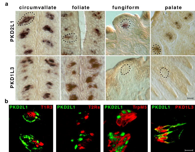
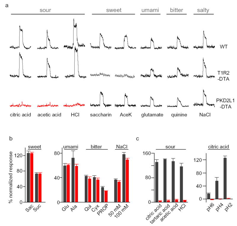
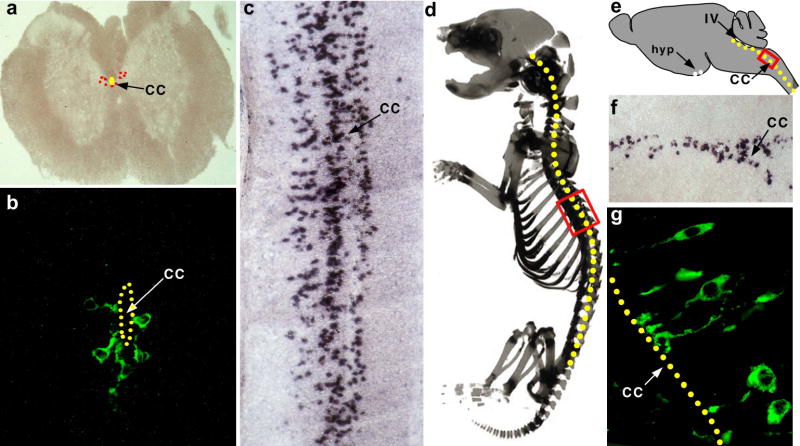
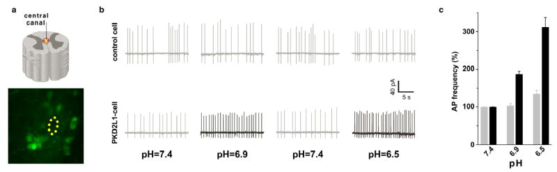
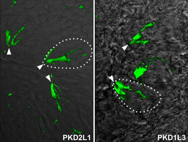
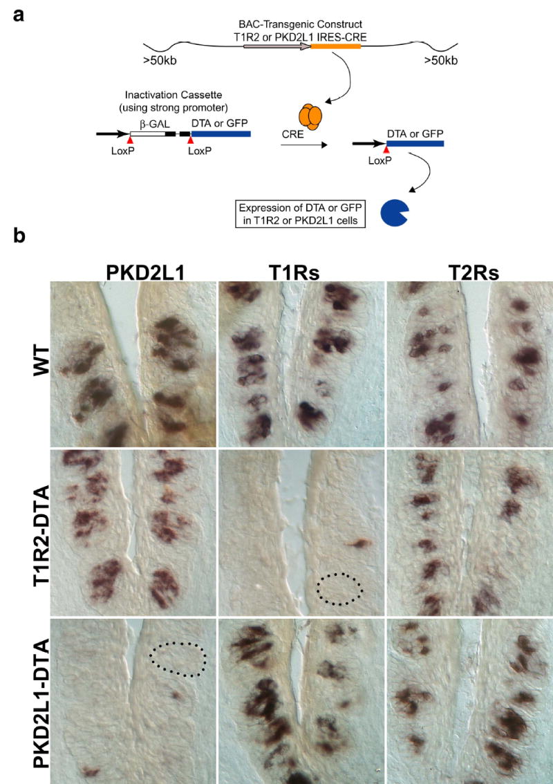
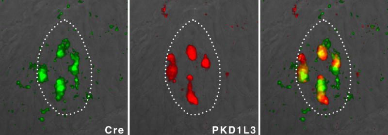
Mammals taste many compounds yet use a sensory palette consisting of only five basic taste modalities: sweet, bitter, sour, salty and umami (the taste of monosodium glutamate). Although this repertoire may seem modest, it provides animals with critical information about the nature and quality of food. Sour taste detection functions as an important sensory input to warn against the ingestion of acidic (for example, spoiled or unripe) food sources. We have used a combination of bioinformatics, genetic and functional studies to identify PKD2L1, a polycystic-kidney-disease-like ion channel, as a candidate mammalian sour taste sensor. In the tongue, PKD2L1 is expressed in a subset of taste receptor cells distinct from those responsible for sweet, bitter and umami taste. To examine the role of PKD2L1-expressing taste cells in vivo, we engineered mice with targeted genetic ablations of selected populations of taste receptor cells. Animals lacking PKD2L1-expressing cells are completely devoid of taste responses to sour stimuli. Notably, responses to all other tastants remained unaffected, proving that the segregation of taste qualities even extends to ionic stimuli. Our results now establish independent cellular substrates for four of the five basic taste modalities, and support a comprehensive labelled-line mode of taste coding at the periphery. Notably, PKD2L1 is also expressed in specific neurons surrounding the central canal of the spinal cord. Here we demonstrate that these PKD2L1-expressing neurons send projections to the central canal, and selectively trigger action potentials in response to decreases in extracellular pH. We propose that these cells correspond to the long-sought components of the cerebrospinal fluid chemosensory system. Taken together, our results suggest a common basis for acid sensing in disparate physiological settings.


Figures

Figure 1
Figure 1. PKD2L1 is expressed in a novel population of TRCs
In situ hybridization (PKD2L1, PKD1L3, T1Rs, T2Rs and TRPM5) and double-label fluorescent immunohistochemistry (PKD2L1) were used to examine the overlap in cellular expression of taste receptors, TRPM5, PKD2L1 and PKD1L3. (a) In situ hybridization of PKD2L1 and PKD1L3 against circumvallate, foliate, fungiform and palate taste buds illustrating expression of PKD2L1 in subsets of TRCs of all taste buds, but lack of PKD1L3 in fungiform and palate TRCs. Dotted lines show the outline of sample taste buds. Scale bar represents 25 μm. (b) The first three panels show co-labeling with a PKD2L1 antisense RNA probe (PKD, green) and T1R3 (T1R, sweet and umami cells), a mixture of 20 T2Rs (bitter cells), and TRPM5 (sweet, umami and bitter cells), respectively. The last panel shows co-labeling with anti-PKD2L1 antibodies and an antisense PKD1L3 RNA probe. Note the absence of overlap between PKD2L1-expressing cells and those expressing sweet, umami or bitter receptors. However, PKD1L3 is always co-expressed with PKD2L1 in CV and foliate papillae. Scale bar represents 10 μm.
Figure 2
Figure 2. PKD2L1-expressing TRCs are the mediators of sour taste
(a) Targeted expression of DTA to selective populations of TRCs produces animals with selective deficits in taste responses. Wild-type mice (WT) show robust neural responses to sour, sweet, umami (amino acid), bitter and salty tastants. However, ablation of sweet cells (T1R2-DTA) generates animals with a dramatic loss of sweet taste (middle panel). In contrast, ablation of PKD2L1-expressing cells eliminates responses to all acid stimuli (bottom panel). Importantly, responses to all other taste qualities remain unimpaired in the DTA-expressing animals. Shown are integrated chorda tympani responses (see Methods). (b) Average neural responses of animals lacking PKD2L1-expressing cells to an expanded panel of tastants; note normal responses to sweet, umami, bitter and salt stimuli. Wild type, gray bars; PKD2L1-DTA, red bars; the values are means ± s.e.m. (n=5) (c) Quantitation of acid responses of wild type and PKD2L1-DTA animals. The values are means ± s.e.m. (n=6). Only the differences in acid responses are significant between wild type and PKD2L1-DTA mice (P<0.00001).
Figure 3
Figure 3. PKD2L1 is expressed in neurons contacting the central canal of the spinal cord
(a) In situ hybridization with PKD2L1 specific probes to a coronal section of the spinal cord; signals are pseudocolored in red. (b) High magnification staining with anti-PKD2L1antibodies reveals a population of PKD2L1-expressing neurons surrounding the central canal of the spinal cord (cc; highlighted by yellow dots in all panels). (c) In situ hybridization with PKD2L1-specific probes on a sagital section of a P1 mouse. The PKD2L1-expressing cells are found throughout the entire length of the spinal cord. (d) Red box denotes the approximate area of the in situ shown in panel (c). (e–f) PKD2L1-expression extends through the brain stem and into the IV ventricle (IV). There is also a very small group of positive cells in the hypothalamus (hyp; data not shown). (g) Immunofluorescent stainings with anti-PKD2L1 antibodies. PKD2L1-expressing neurons project into the central canal; note robust expression of PKD2L1 receptors at the terminals.
Figure 4
Figure 4. PKD2L1-expressing neurons of the central canal fire action potentials in response to pH stimulation
Spinal cord neurons were patched using a loose patch configuration, tested for the presence of basal activity and recorded in the cell-attached configuration. (a) GFP-expressing (PKD2L1-positve cells) or unlabeled (control) cells were examined for pH responses. (b) Responses of a sample GFP-labeled or unlabeled neuron to test solutions under a perfusion regime consisting of pH 7.4, pH 6.9, pH 7.4 and pH 6.5; shown are AP traces in a window of ~25 sec. (c) Data were analyzed by examining records of ~4 minutes at each pH condition. Basal activity ranged between 1–5 Hz. Note the dramatic increases in pH-evoked firing frequency in GFP-labeled neurons versus unlabeled cells (P<0.001). A minimum of 8 GFP-labeled and 5 unlabelled cells were characterized for each stimulus. The values are means + s.e.m. normalized to basal activity at pH 7.4 (taken as 100%).
Supplementary Figure 1
PKD2L1 and PKD1L3 are enriched in the taste pore Immunofluorescent stainings of mouse taste buds with PKD2L1 (left panel) and with PKD1L3 (right panel) antibodies. The pictures show superposition of fluorescent antibody signals on DIC images of taste tissue. Dotted lines illustrate the outline of a taste bud, and arrows point to the taste pore region
Supplementary Figure 2
Loss of selective TRCs in DTA-expressing animals Upper diagram illustrates the strategy used to target DTA or GFP to selective populations of TRCs. BAC constructs contained the entire T1R2 or PKD2L1 genes with the IRES-Cre added downstream of the termination codon, but upstream of polyA-addition signals. In both cases, the transgenic constructs included at least 50Kb of flanking sequences upstream and downstream of the target gene (see Methods). Fidelity of Cre and reporter expression in the correct cell types was confirmed by double labeling with a variety of TRC-specific gene probes. Lower panels show in situ hybridization experiments examining the presence of sweet (T1Rs), bitter (T2Rs) or PKD2L1-expressing cells in the two engineered lines. Targeting of DTA to T1R2- or PKD2L1-expressing cells eliminates over 95% of their respective TRC population. In situ hybridization probes were as in Figure 1.
Supplementary Figure 3
Targeting of Cre recombinase to PKD-expressing TRCs In situ hybridization with double-labeled probes (Cre and PKD1L3) was used to examine the expression of Cre recombinase in PKD-expressing cells. Dotted lines illustrate the outline of a taste bud; note the cellular overlap in the hybridization signals. Similar results were obtained by crossing PKD2L1-Cre lines to GFP reporter lines.

References

    1. Lindemann B. Receptors and transduction in taste. Nature. 2001;413:219–25. - PubMed
    1. Kinnamon SC, Margolskee RF. Mechanisms of taste transduction. Curr Opin Neurobiol. 1996;6:506–13. - PubMed
    1. DeSimone JA, Lyall V, Heck GL, Feldman GM. Acid detection by taste receptor cells. Respir Physiol. 2001;129:231–45. - PubMed
    1. Wu G, et al. Identification of PKD2L, a human PKD2-related gene: tissue-specific expression and mapping to chromosome 10q25. Genomics. 1998;54:564–8. - PubMed
    1. Adler E, et al. A novel family of mammalian taste receptors. Cell. 2000;100:693–702. - PubMed

Publication types

MeSH terms