. 2006 Jun 12:1:2.
doi: 10.1186/1750-1326-1-2.

Presenilin-dependent intramembrane cleavage of ephrin-B1

Affiliations

Presenilin-dependent intramembrane cleavage of ephrin-B1

Taisuke Tomita et al. Mol Neurodegener. .

Abstract

Background: Presenilin-dependent gamma-secretase cleavage of several transmembrane proteins, including amyloid-beta precursor protein and Notch, mediates the intramembrane proteolysis to liberate their intracellular domains that are involved in cellular signaling. Considering gamma-secretase inhibitors as therapeutics for Alzheimer's disease, understanding the physiologically and biologically important substrate for gamma-secretase activity in brains is emerging issue. To elucidate the molecular mechanism and physiological role of gamma-secretase, we screened candidate molecules for gamma-secretase substrates.

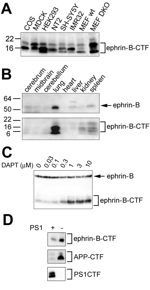
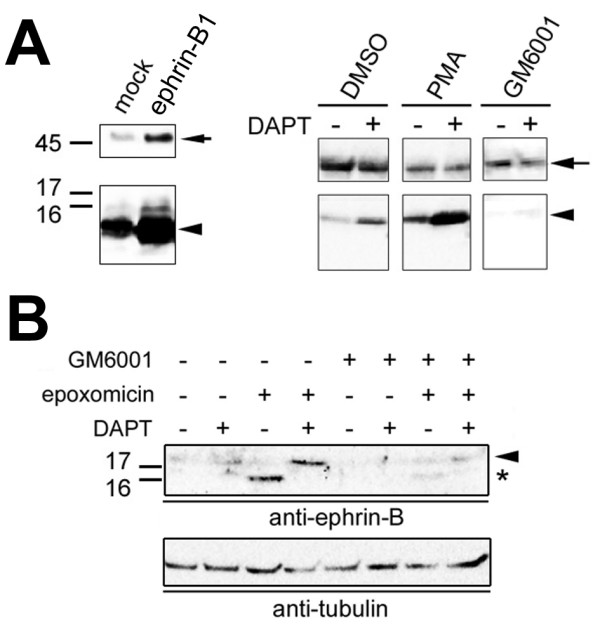
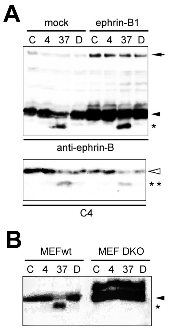
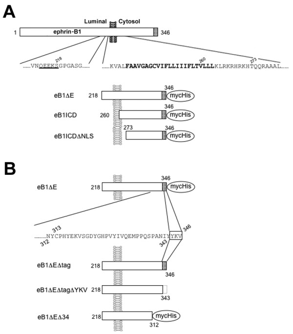
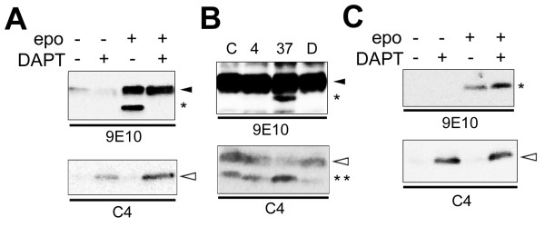
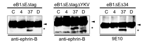
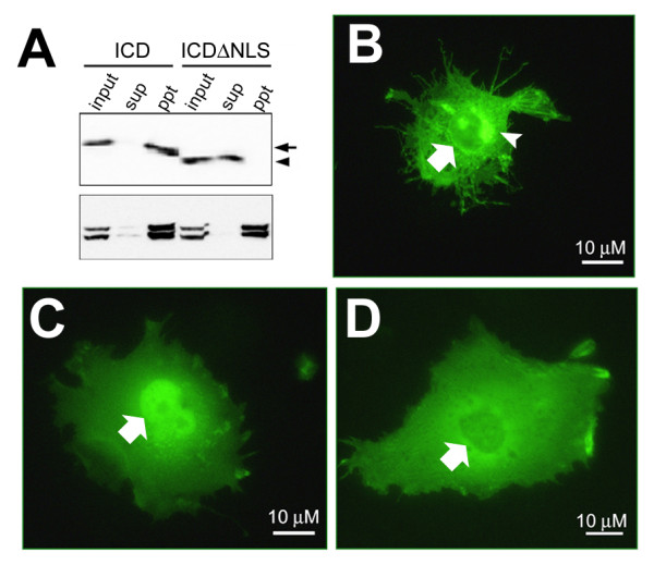
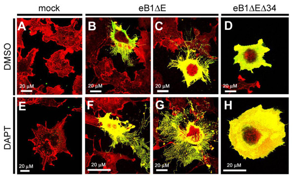
Results: We show that ephrin-B1, that participates in cell-cell repulsive and attractive signaling together with its Eph receptor, constitutively undergoes ectodomain shedding and that the residual membrane-tethered fragment is sequentially cleaved by gamma-secretase to release the intracellular domain. Furthermore, overexpression of membrane-tethered ephrin-B1 caused protrusion of numerous cellular processes consisted of F-actin, that required the preservation of the most C-terminal region of ephrin-B1. In contrast, soluble intracellular domain translocated into the nucleus and had no effect on cell morphology.

Conclusion: Our findings suggest that ephrin-B is a genuine substrate for gamma-secretase and regulates the cytoskeletal dynamics through intramembrane proteolysis.


Figures

Figure 1
Figure 1
Immunoblot analysis of endogenous ephrin-B CTFs. Membrane fractions from cultured cells (A) and mouse organs (B) were subjected to immunoblot analyses using anti-ephrin-B antibody. C) Immunoblot analysis of lysates from COS cells treated with a γ-secretase inhibitor DAPT. D) Immunoblot analysis of lysates from DKO cells expressing PS1.
Figure 2
Figure 2
Analysis of proteolytic processing of ephrin-B1. A) Immunoblot analysis of COS cells overexpressing ephrin-B1 (arrows). ephrin-B1-CTF (arrowheads) was increased by DAPT and PMA treatment, whereas GM6001 completely diminished the ephrin-B1-CTF. B) Effect of protease inhibitors on endogenous ephrin-B CTF. Note that proteasome inhibition by epoxomicin caused an accumulation of CTF (arrowhead) and ICD (asterisk), that was abolished by GM6001 treatment.
Figure 3
Figure 3
Cell-free γ-secretase assay for ephrin-B1 cleavage. A) Cell-assay using membranes from mock- or ephrin-B1-transfected COS cells. Samples incubated at 4 degree (4), 37 degree (37) or 37 degree with DAPT (D) were analyzed by immunoblotting by anti-ephrin-B antibody (Upper panel). Same fractions without incubation were indicated as "C". ephrin-B1 FL, ephrin-B-CTF and de novo generated ICD were shown by black arrow, arrowhead and asterisk, respectively. Endogenous APP (Lower panel) C-terminal stubs (white arrowhead) were cleaved by γ-secretase to generate AICD (double asterisks). B) Cell-free assay of membranes from wild-type MEF (MEFwt) and MEF lacking Psen1 and Psen2 (DKO). ephrin-B-CTF and de novo generated ICD were indicated by arrowhead and asterisk.
Figure 4
Figure 4
Schematic depiction of ephrin-B1 derivatives used in this study. A) N-terminally truncated ephrin-B1 derivatives. B) C-terminally truncated eB1ΔE derivatives. Putative amino acid sequence recognized by sheddase is underlined. Transmembrane domain is shown in bold. PDZ domain binding region (YKV) and Myc/His tag are indicated by shaded box and oval, respectively. Numbers indicate the residues in ephrin-B1 FL protein.
Figure 5
Figure 5
Proteolytic processing of overexpressed ephrin-B1 derivatives. A) Proteasome and γ-secretase inhibitor ("epo" and "DAPT", respectively) treatments on cells expressing eB1ΔE (black arrowhead). Proteolytically-generated ICD was shown by asterisk. DAPT inhibited the processing of eB1ΔE as well as endogenous APP (accumulation of C-terminal stub, white arrowhead in lower panel). B) Cell-free assay using COS membrane expressing eB1ΔE. de novo generated ICDs from eB1ΔE (black arrowhead) and APP C-terminal stub (white arrowhead) were indicated by asterisk and double asterisks, respectively. C) Inhibitor treatments (same as A) on cells expressing eB1ICD (asterisk). Accumulation of C-terminal stub (white arrowhead) by DAPT was shown at lower panel.
Figure 6
Figure 6
Proteolytic processing of overexpressed eB1ΔE derivatives. Cell-free assay using COS membranes expressing eB1ΔEΔtag (left), eB1ΔEΔtagΔYKV (middle) and eB1ΔEΔ34 (right). Antibodies used were indicated below the panels. All derivatives (arrowheads) were cleaved to generate ICD-like peptides (asterisks) by γ-secretase activity.
Figure 7
Figure 7
eB1ICD translocated into the nucleus. A) Immunoblot analysis of total (input), Triton-soluble (sup) or -insoluble (ppt) lysates from COS cells expressing eB1ICD (arrow) or eB1ICDΔNLS (arrowhead) using anti-myc (upper panel) or anti-lamin A/C (lower panel). B), C) and D) were the representative results of immunocytochemical analysis of COS cells expressing eB1ΔE, eB1ICD and eB1ICDΔNLS, respectively, using anti-myc antibody. Nucleus and golgi area were indicated by arrows and arrowhead, respectively.
Figure 8
Figure 8
Immunocytochemical analysis of COS cells expressing eB1ΔE and eB1ΔEΔ34. COS cells were transfected with mock (A, E), eB1ΔE (B, C, F, G) or eB1ΔEΔ34 (D, H) and treated with DMSO (A-D) or DAPT (E-H). Fixed cells were probed with anti-myc (Green) and Rhodamin-Phalloidin (Red) for eB1ΔE and F-actin, respectively.

References

    1. Tomita T, Iwatsubo T. The inhibition of γ-secretase as a therapeutic approach to Alzheimer's disease. Drug News Perspect. 2004;17:321–325. doi: 10.1358/dnp.2004.17.5.829036. - DOI - PubMed
    1. Takasugi N, Tomita T, Hayashi I, Tsuruoka M, Niimura M, Takahashi Y, Thinakaran G, Iwatsubo T. The role of presenilin cofactors in the γ-secretase complex. Nature. 2003;422:438–441. doi: 10.1038/nature01506. - DOI - PubMed
    1. Kopan R, Ilagan MX. γ-Secretase: proteasome of the membrane? Nat Rev Mol Cell. 2004;5:499–504. doi: 10.1038/nrm1406. - DOI - PubMed
    1. De Strooper B, Annaert W, Cupers P, Saftig P, Craessaerts K, Mumm JS, Schroeter EH, Schrijvers V, Wolfe MS, Ray WJ, Goate A, Kopan R. A presenilin-1-dependent γ-secretase-like protease mediates release of Notch intracellular domain. Nature. 1999;398:518–522. doi: 10.1038/19083. - DOI - PubMed
    1. Landman N, Kim TW. Got RIP? Presenilin-dependent intramembrane proteolysis in growth factor receptor signaling. Cytokine Growth Factor Rev. 2004;15:337–351. doi: 10.1016/j.cytogfr.2004.04.001. - DOI - PubMed

LinkOut - more resources