. 2006 Aug 24:7:32.
doi: 10.1186/1471-2121-7-32.

The fission yeast DNA structure checkpoint protein Rad26ATRIP/LCD1/UVSD accumulates in the cytoplasm following microtubule destabilization

Affiliations

The fission yeast DNA structure checkpoint protein Rad26ATRIP/LCD1/UVSD accumulates in the cytoplasm following microtubule destabilization

Erin E Baschal et al. BMC Cell Biol. .

Abstract

Background: DNA structure checkpoints are conserved eukaryotic signal transduction pathways that help preserve genomic integrity. Upon detecting checkpoint signals such as stalled replication forks or double-stranded DNA breaks, these pathways coordinate appropriate stress responses. Members of the PI-3 kinase related kinase (PIKK) family are essential elements of DNA structure checkpoints. In fission yeast, the Rad3 PIKK and its regulatory subunit Rad26 coordinate the detection of checkpoint signals with pathway outputs.

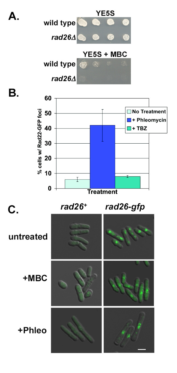
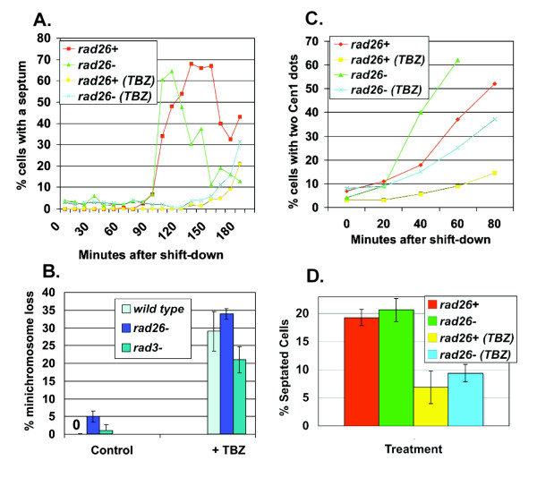
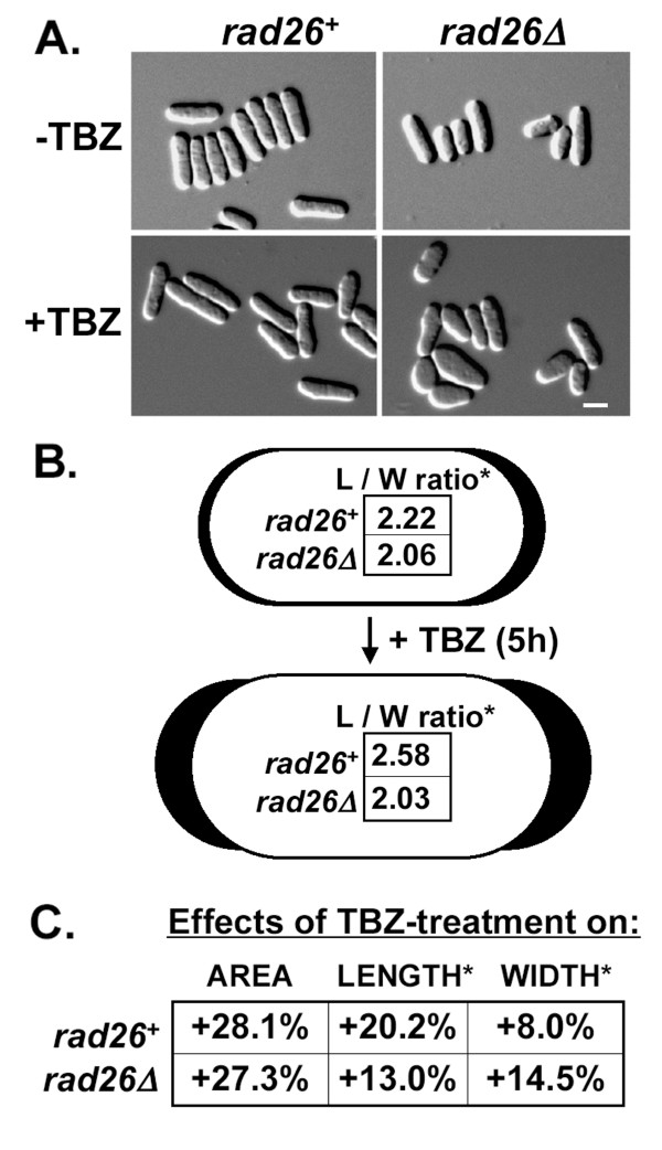
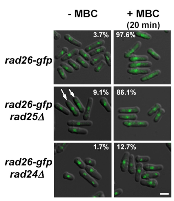
Results: We found that untreated rad26Delta cells were defective for two microtubule-dependent processes: chromosome segregation and morphogenesis. Interestingly, cytoplasmic accumulation of Rad26-GFP occurred following treatment with microtubule destabilizing drugs, but not during treatment with the genotoxic agent Phleomycin. Cytoplasmic accumulation of Rad26-GFP depended on Rad24, a 14-3-3 protein also required for DNA structure checkpoints and morphogenesis. Results of over expression and epistasis experiments confirm that Rad26 and Rad24 define a response to microtubule destabilizing conditions.

Conclusion: Two DNA structure checkpoint proteins with roles in morphogenesis define a response to microtubule destabilizing conditions.


Figures

Figure 1
Figure 1
Rad26 responds to conditions that destabilize microtubules. A. rad26Δ cells were sensitive to 8 μg/ml MBC. Cultures of TE236 (rad26+) and TE257 (rad26Δ) were serially diluted onto YE5S and YE5S + MBC plates and grown for 4 days at 30°C. B. TBZ did not cause relocalization of Rad22-GFP. A culture of TE1239 (rad22-gfp) was split and then treated with 20 μg/ml TBZ for five hours, 7.5 μg/ml Phleomycin for two hours, or left untreated. Bars = Std dev C. Rad26-GFP accumulated in the cytoplasm following MBC, but not Phleomycin, treatment. TE236 (rad26+) and TE1197 (rad26-gfp) cells cultured in minimal medium (EMM) were left untreated or treated with 8 μg/ml MBC for 3 hours or 10 μg/ml Phleomycin for 4 hours, fixed with paraformaldehyde and processed for microscopy. The Rad26-GFP signal was similar in live cells (data not shown). Bar = 7 μm.
Figure 2
Figure 2
The spindle assembly checkpoint of rad26Δ cells appears functional. A. rad26Δ cells delayed mitosis during TBZ treatment. Cultures of TW1261 (cdc25.22 rad26+) and TW1262 (cdc25.22 rad26Δ) were shifted to 37°C for 4 hours to arrest cells in G2. TBZ (20 μg/ml) was added to respective cultures 30 minutes before shifting to 30°C and releasing into mitosis. Cells were fixed with paraformaldehyde and stained with Calcofluor to visualize septa. B. Chromosome stability was not affected in TBZ-treated rad26Δ cells. The adenine-marked minichromosome of TE787 (rad3Δ), TW1222 (wild type) and TW1224 (rad26Δ) was used to assay chromosome loss following 8 h of TBZ treatment. Bars = Std dev C. Chromosome separation was restrained in rad26Δ cells during TBZ treatment. Cultures of TW1261 and TW1262 were shifted to 37°C for 4 hours and arrested in G2. TBZ was added to respective cultures 30 minutes before shifting to 26°C and releasing into mitosis. Cells were fixed in methanol; chromosome separation was monitored using the Cen1-GFP marker. D. The septation index of asynchronous rad26Δ cultures is elevated. Asynchronous cultures of rad26+ (TE236) and rad26Δ (TE257) cells were split and left untreated or treated with 20 μg/ml TBZ for five hours, fixed with paraformaldehyde and stained with Calcofluor. The septation index is the percentage of septated cells in the culture.
Figure 3
Figure 3
TBZ affected the polarity of rad26Δ cells. A. Images of untreated and TBZ-treated rad26+ and rad26Δ cells. Cultures of rad26+ (TE236) and rad26Δ (TE257) cells were split and left untreated or treated with 20 μg/ml TBZ for five hours, fixed with paraformaldehyde and observed using DIC. B. Interpretive diagram showing that rad26Δ cells were significantly wider (*) than rad26+ cells. The dimensions of untreated and TBZ-treated cells were quantified using Leica FW4000 image analysis software. The length-to-width (L/W) ratios were calculated and presented here in pictorial representations of rad26+ (outlined in black) and rad26Δ (outlined in white) cells. C. The width of rad26Δ cells increased (+) during TBZ-treatment.
Figure 4
Figure 4
Additional polarity defects associated with loss or over expression of rad26+. A. – D. Prolonged G2 arrest affected the morphology of rad26Δ cells. A. rad26+ cdc25.22 (TW1261) and B. to D. rad26Δ cdc25.22 (TW1262) cells were arrested at 37°C for 3 h, fixed and stained with Calcofluor. Bar = 8 μm. E. and F. rad26Δ exacerbated the polarity defects of kin1Δ cells. E. kin1Δ (TE550) and F. kin1Δ rad26Δ were grown in liquid culture, fixed with paraformaldehyde and observed with brightfield. Bar = 10 μm. G. – I. Over expression (OE) of rad26+ caused polarity defects. G. rad26+ with empty vector (TE236 with pTE102), H. rad26+ OE rad26+ (TE236 with pTE169) and I. rad3Δ OE rad26+ (TE570 with pTE169) cells were grown in promoter-derepressing conditions for 20 hours, fixed with paraformaldehyde and stained with DAPI. The arrow points to a cell with an abnormal number of nuclei. Bar = 10 μm.
Figure 5
Figure 5
Loss of rad26+ affected the polar distribution of actin patches but not gross microtubule architecture. A. Gross microtubule architecture was not affected by loss of rad26+. rad26+ (1226) and rad26Δ (1248) cells expressing ectopic atb2-gfp were grown in EMM + 0.2 μM thiamine. TBZ (20 μg/ml) was added to half of each culture for 5 hours, after which cells were fixed with methanol and processed for microscopy. Bar = 4 μm B. rad26+ is required for the polar distribution of actin patches. rad26+ (TE236) and rad26Δ (TE257) cells were grown in YE5S liquid. Half of each culture was treated with 20 μg/ml TBZ for 5 hours before fixing and staining with FITC-Phalloidin (see Methods). Bar = 5 μm C. A diagrammatic representation of polar and non-polar actin patch distributions. D. Graphical representation of data collected from B.
Figure 6
Figure 6
Over expression of rad24 cDNA rescued the TBZ-sensitivity of rad26Δ cells, but not mad2Δ cells. A. Results of the over expression (OE) screen (see Methods). Four cDNAs suppressed the TBZ-sensitivity of rad26Δ cells, and three of these also suppressed the TBZ-sensitivities of nda2KM52 and mad2Δ cells. B. Only OErad24+ specifically suppressed the growth of rad26Δ cells on TBZ. Wild type (TE236), rad26Δ (TE257) and mad2Δ (TW1219) were transformed with a plasmid containing inducible rad24 cDNA (pTW909) and streaked onto EMM and EMM + TBZ medium containing the vital stain Phloxin B. Pictures were taken after 3 days of growth at 30°C. Upper left = EMM; Upper right = EMM + TBZ (20 μg/ml) C. rad24Δ cells were sensitive to TBZ. Wild type (TE236), rad26Δ (TE257), rad24Δ (TE465), rad25Δ (TE464) and chk1Δ cds1Δ (TE892) were streaked onto YE5S (top) and YE5S + 20 μg/ml TBZ (bottom) and incubated at 30°C for three days.
Figure 7
Figure 7
The rad26Δ and rad24Δ alleles may cause TBZ-sensitivity by a pathway that is independent of the cytokinesis checkpoint. A. The rad26Δ rad24Δ strain did not display an additive phenotype on TBZ medium. Cultures of wild type (TE236), rad26Δ (TE257), rad24Δ (TE465) and rad26Δ rad24Δ (TW1235) were serially diluted and manually spotted onto YE5S and YE5S + 8, 14 and 16 μg/ml TBZ. Pictures were taken after 3 days of growth at 30°C. B. rad24Δ, but not rad26Δ, was sensitive to LatA. Cultures were spotted onto YE5S plates + 0.5 μM LatA. C. The cytokinesis checkpoint of rad26Δ cells was intact. Liquid YE5S cultures of wild type (TE236), rad26Δ (TE257) and rad24Δ (TE465) were left untreated or treated with 0.2 μM LatA for 5 hours, fixed with paraformaldehyde and stained with DAPI.
Figure 8
Figure 8
rad24+ was required for normal cytoplasmic accumulation of Rad26-GFP after 20 minutes of MBC treatment. Cultures of rad26-gfp (TE1197), rad25Δ rad26-gfp (TW1237) and rad24Δ rad26-gfp (TW1238) in liquid EMM minimal media were left untreated or treated with MBC for 20 minutes. The figure was made by merging DIC and GFP images. Arrows point to untreated rad25Δ rad26-gfp cells that have cytoplasmic Rad26-GFP signal. Notice that rad24Δ cells are more spherical than rad24+ and rad25Δ cells. The percentage of cells containing cytoplasmic Rad26-GFP signal is shown (N > 100). Bar = 5 μm.

Similar articles

Cited by

References

    1. Carr AM. DNA structure dependent checkpoints as regulators of DNA repair. DNA Repair (Amst) 2002;1:983–994. - PubMed
    1. d'Adda di Fagagna F, Reaper PM, Clay-Farrace L, Fiegler H, Carr P, Von Zglinicki T, Saretzki G, Carter NP, Jackson SP. A DNA damage checkpoint response in telomere-initiated senescence. Nature. 2003;426:194–198. - PubMed
    1. Bakkenist CJ, Kastan MB. Initiating cellular stress responses. Cell. 2004;118:9–17. - PubMed
    1. Bentley NJ, Holtzman DA, Flaggs G, Keegan KS, DeMaggio A, Ford JC, Hoekstra M, Carr AM. The Schizosaccharomyces pombe rad3 checkpoint gene. EMBO J. 1996;15:6641–6651. - PMC - PubMed
    1. Edwards RJ, Bentley NJ, Carr AM. A Rad3-Rad26 complex responds to DNA damage independently of other checkpoint proteins. Nat Cell Biol. 1999;1:393–398. - PubMed

Publication types

MeSH terms