

The frequency of B- and T-cell gene rearrangements and epstein-barr virus in T-cell lymphomas: a comparison between angioimmunoblastic T-cell lymphoma and peripheral T-cell lymphoma, unspecified with and without associated B-cell proliferations
- PMID: 16931587
- PMCID: PMC1867616
- DOI: 10.2353/jmoldx.2006.060016
The frequency of B- and T-cell gene rearrangements and epstein-barr virus in T-cell lymphomas: a comparison between angioimmunoblastic T-cell lymphoma and peripheral T-cell lymphoma, unspecified with and without associated B-cell proliferations
Abstract
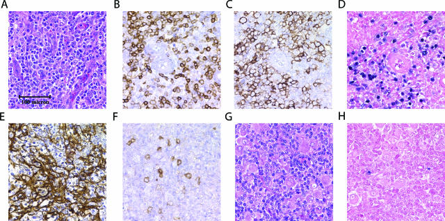
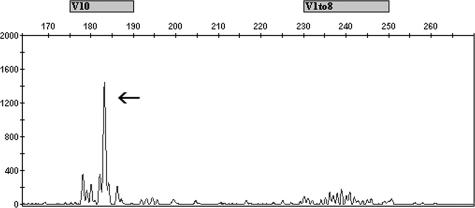
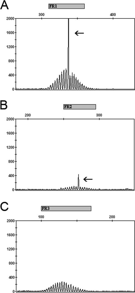
We report on a series of 58 cases of angioimmunoblastic T-cell lymphoma (AILT) and 59 cases of peripheral T-cell lymphoma, unspecified (PTCL-NOS). Subsets of cases from both diagnostic groups were complicated by associated B-cell proliferations, and we performed B- and T-cell clonality studies and in situ hybridization for Epstein-Barr virus (EBV) to investigate the relationship between B-cell proliferation, B-cell clonality, and EBV. Using multiplex polymerase chain reaction assays based on the BIOMED-2 collaborative study, we detected TCRgamma T-cell clones in 78 and 81% of AILT and PTCL-NOS cases, respectively, and IGH B-cell clones in 34 and 35% of AILT and PTCL-NOS cases, respectively. The majority of cases contained EBV-positive cells, including 50% of AILT and 57% of PTCL-NOS cases, and cases with B-cell proliferations were more often EBV-positive. Although a relatively high rate of B-cell clonality has been shown for AILT, our findings for PTCL-NOS differ from previous reports in that B-cell clonality was relatively frequent. Overall, a positive B-cell clone correlated, in part, with the presence of a B-cell proliferation but not with EBV. Our findings demonstrate that B-cell clonality is a common finding in AILT and PTCL-NOS, and its presence should not negate the diagnosis established by morphologic, immunophenotypic, and clinical findings.
Figures
Comment in
-
Immunoglobulin and T-cell receptor gene rearrangements: minding your B's and T's in assessing lineage and clonality in neoplastic lymphoproliferative disorders.J Mol Diagn. 2006 Sep;8(4):426-9; quiz 526-7. doi: 10.2353/jmoldx.2006.060108. J Mol Diagn. 2006. PMID: 16931581 Free PMC article. No abstract available.
References
-
- Jaffe ES, Harris NL, Stein H, Vardiman JW. Kleihues P, Sobin LH, editors. Lyon: IARC Press,; Pathology and Genetics of Tumours of Haematopoietic and Lymphoid Tissues. 2001
-
- Theriault C, Galoin S, Valmary S, Selves J, Lamant L, Roda D, Rigal-Huguet F, Brousset P, Delsol G, Al Saati T. PCR analysis of immunoglobulin heavy chain (IgH) and TcR-gamma chain gene rearrangements in the diagnosis of lymphoproliferative disorders: results of a study of 525 cases. Mod Pathol. 2000;13:1269–1279. - PubMed
-
- Garcia MJ, Martinez-Delgado B, Granizo JJ, Benitez J, Rivas C. IgH, TCR-gamma, and TCR-beta gene rearrangement in 80 B- and T-cell non-Hodgkin’s lymphomas: study of the association between proliferation and the so-called “aberrant” patterns. Diagn Mol Pathol. 2001;10:69–77. - PubMed
-
- Vergier B, Dubus P, Kutschmar A, Parrens M, Ferrer J, de Mascarel A, Merlio JP. Combined analysis of T cell receptor gamma and immunoglobulin heavy chain gene rearrangements at the single-cell level in lymphomas with dual genotype. J Pathol. 2002;198:171–180. - PubMed
Publication types
MeSH terms
Substances
LinkOut - more resources
Full Text Sources
