. 2006 Aug 25;313(5790):1126-30.
doi: 10.1126/science.1127119.

Symbiotic bacteria direct expression of an intestinal bactericidal lectin

Affiliations

Symbiotic bacteria direct expression of an intestinal bactericidal lectin

Heather L Cash et al. Science. .

Abstract

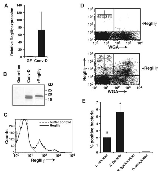
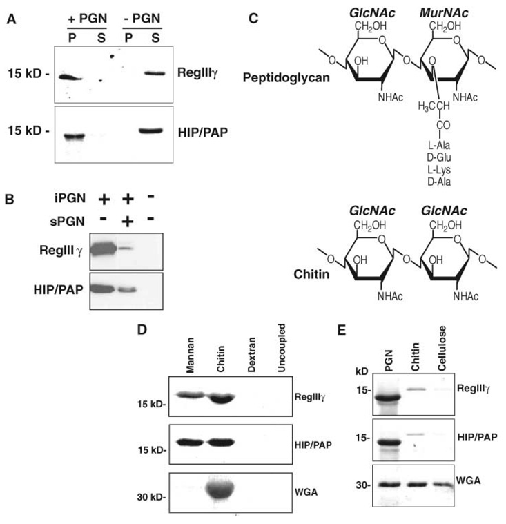
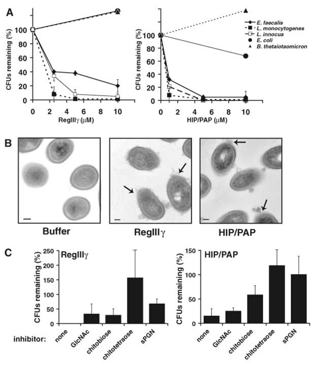
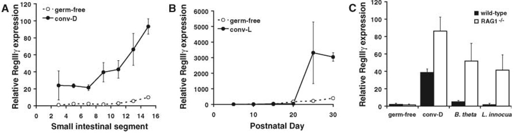
The mammalian intestine harbors complex societies of beneficial bacteria that are maintained in the lumen with minimal penetration of mucosal surfaces. Microbial colonization of germ-free mice triggers epithelial expression of RegIIIgamma, a secreted C-type lectin. RegIIIgamma binds intestinal bacteria but lacks the complement recruitment domains present in other microbe-binding mammalian C-type lectins. We show that RegIIIgamma and its human counterpart, HIP/PAP, are directly antimicrobial proteins that bind their bacterial targets via interactions with peptidoglycan carbohydrate. We propose that these proteins represent an evolutionarily primitive form of lectin-mediated innate immunity, and that they reveal intestinal strategies for maintaining symbiotic host-microbial relationships.


Figures

Fig. 1
Fig. 1
RegIIIγ is induced by resident intestinal microbes and binds to Gram-positive bacteria. (A) RegIIIγ mRNA expression in Paneth cells. Paneth cells were harvested by laser-capture microdissection from germ-free and conventionalized small intestines. Q-PCR analysis was performed on RNAs from microdissected Paneth cells from three mice per group. Values were normalized to glyceraldehyde-3-phosphate dehydrogenase (GAPDH) expression, and mean ± SD is plotted (range for conventionalized samples is 42 to 309). Results are expressed relative to one of the germ-free samples. GF, germ-free; Conv-D, conventionalized. (B) RegIIIγ protein expression in small intestine. RegIIIγ was detected in mid–small intestinal protein by immunoblot with RegIIIγ-specific antiserum (13). The lower band in the protein sample from conventionalized mice likely results from proteolytic cleavage at a trypsin-like site near the N-terminus (13), similar to that described for RegIα (8). Results are representative of two independent experiments. rRegIIIγ, recombinant RegIIIγ. (C) RegIIIγ binds to intestinal bacteria. Flow cytometry reveals binding of AlexaFluor555–conjugated RegIIIγ to intestinal bacteria recovered from conventional mouse small intestine. BSA-AlexaFluor555 showed no binding (not shown). Results are representative of independent experiments with three mice. (D) RegIIIγ binds preferentially to Gram-positive intestinal bacteria. Dual-color flow cytometry analysis with WGA-AlexaFluor488 and RegIIIγ-AlexaFluor555 shows preferential binding to the WGA-positive bacterial population. Results are representative of three independent experiments. (E) RegIIIγ binds preferentially to cultured Gram-positive bacteria. Formaldehyde-fixed preparations of Gram-positive (L. innocua and E. faecalis) and Gram-negative bacteria (S. typhimurium and P. aeruginosa) were incubated with RegIIIγ, followed by detection with RegIIIγ-specific antiserum and Cy3-labeled goat secondary antibody against rabbit IgG, and analyzed by flow cytometry. Results are representative of three experiments. Asterisks indicate statistically significant differences between Gram-positive species and S. typhimurium (P < 0.05). Preimmune serum controls are shown in fig. S3.
Fig. 2
Fig. 2
Mouse RegIIIγ and human HIP/PAP bind peptidoglycan. (A) Peptidoglycan pull-down assays. RegIIIγ or HIP/PAP (10 µg of either) was added to 50 µg insoluble Bacillus subtilis peptidoglycan and pelleted. Pellet (P) and supernatant (S) fractions were analyzed by SDS–polyacrylamide gel electrophoresis (SDS-PAGE) and Coomassie blue staining. (B) sPGN competes with iPGN for lectin binding. Pull-down assays were performed with or without 100 µM soluble B. subtilis peptidoglycan. (C) Comparison of peptidoglycan and chitin structures. The structure of a typical Gram-positive peptidoglycan is depicted. (D) Lectin binding to immobilized polysaccharides. Lectins were bound to immobilized polysaccharide for 2 hours at 4°C. After washing, bound proteins were released by boiling in SDS-PAGE sample buffer and analyzed by SDS-PAGE and Coomassie blue staining. (E) Pull-down assays comparing binding to peptidoglycan, chitin, and cellulose. Purified recombinant RegIIIγ or HIP/PAP (10 µg of either) was added to 50 µg of peptidoglycan, chitin, or cellulose and analyzed as in (A). The lower molecular weight forms of RegIIIγ and HIP/PAP in (A) and (E) result from cleavage at an N-terminal trypsinlike site by a peptidoglycan-associated proteolytic activity.
Fig. 3
Fig. 3
Mouse RegIIIγ and human HIP/PAP have antibacterial activity against Gram-positive bacteria. (A) Percentage of CFUs remaining after exposure to purified RegIIIγ and HIP/PAP. L innocua, L monocytogenes, E. faecalis, Escherichia coli K12, and B. thetaiotaomicron were grown to mid–log phase and incubated with purified lectins. Initial bacterial concentrations ranged from 105 to 106 CFU/ml. After incubation for 2 hours at 37°C, viable bacteria were quantified by dilution plating. Assays were done in triplicate. Mean ± SD is plotted. (B) Transmission electron microscopy of L. monocytogenes following a 2-hour exposure to 10 µM purified recombinant RegIIIγ and HIP/PAP. Arrows indicate examples of damaged cell surfaces and cytoplasmic leakage. Scale bar, 100 nm. (C) Lectin bactericidal activity is inhibited by chitooligosaccharides and sPGN. GlcNAc, chitobiose (GlcNAc2), or chitotetraose (GlcNAc4) at 10 mM or 35 µM sPGN was added to antibacterial assays performed on L. innocua as in (A). Each percentage CFU was calculated relative to a no-lectin control assay containing an identical amount of chitooligosaccharide or sPGN.
Fig. 4
Fig. 4
RegIIIγ expression is triggered by intestinal bacteria. (A) RegIIIγ expression along the cephalocaudal axis of the small intestine. Small intestines from adult germ-free or conventionalized (conv-D) NMRI mice were divided into 16 equal segments (numbered proximal to distal) and RegIIIγ mRNA expression was determined in specific segments using Q-PCR. Results are representative of experiments in two sets of mice. (B) RegIIIγ mRNA increases during the weaning period (P17 to 22) in developing conventionally raised NMRI mice. Assays were performed on pooled mid–small intestinal RNAs (for three mice per time point). (C) RegIIIγ expression is triggered by single Gram-positive or Gram-negative bacterial species in immunodeficient mice. Q-PCR determinations were done on cDNAs from mid–small intestine. Each point represents the average value from three or more different mice. All Q-PCR determinations were performed in triplicate (mean ± SD plotted) and were normalized to 18S ribosomal RNA.

Comment in

References

    1. Hooper LV, Midtvedt T, Gordon JI. Annu. Rev. Nutr. 2002;22:283. - PubMed
    1. Wehkamp J, et al. Gut. 2004;53:1658. - PMC - PubMed
    1. Wehkamp J, et al. Proc. Natl. Acad. Sci. U.S.A. 2005;102:18129. - PMC - PubMed
    1. Christa L, et al. Am. J. Physiol. 1996;271:G993. - PubMed
    1. Ogawa H, et al. Inflamm. Bowel Dis. 2003;9:162. - PubMed

Publication types

MeSH terms

Associated data