Review
. 2006 Feb;2(2):99-108.
doi: 10.1038/ncpendmet0098.

Technology insight: In vitro culture of spermatogonial stem cells and their potential therapeutic uses

Affiliations
Review

Technology insight: In vitro culture of spermatogonial stem cells and their potential therapeutic uses

Hiroshi Kubota et al. Nat Clin Pract Endocrinol Metab. 2006 Feb.

Abstract

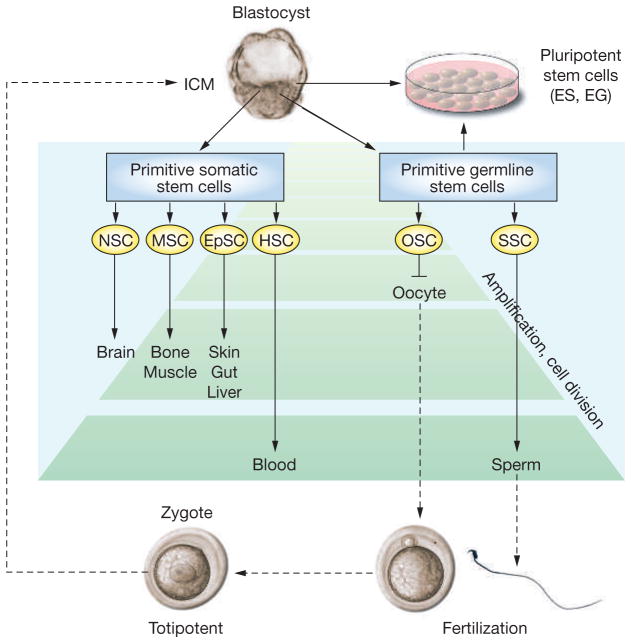
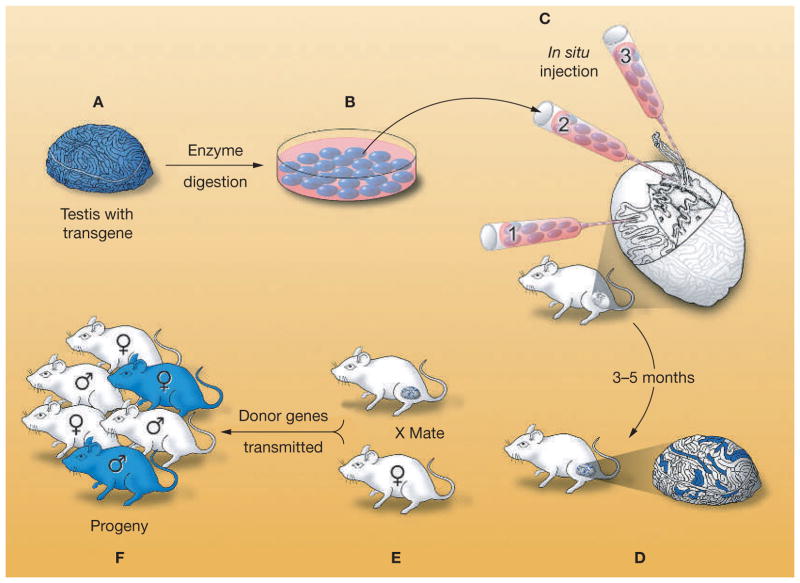
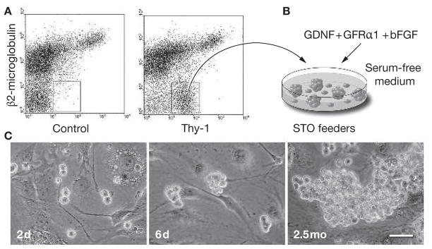
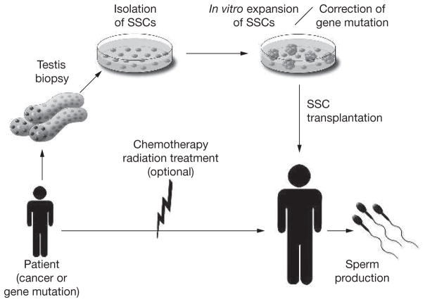
Male germline stem cells--spermatogonial stem cells (SSCs)--self-renew and produce large numbers of differentiating germ cells that become spermatozoa throughout postnatal life and transmit genetic information to the next generation. SSCs are the only germline stem cells in adults, because all female germline stem cells cease proliferation before birth. In this article, we first summarize development of SSCs, and then the relation of SSCs to somatic stem cells in tissues and pluripotent stem cells in vitro, such as embryonic stem cells. Next, we describe a transplantation technique in which donor testis cells from a fertile male can be transplanted to the testes of an infertile male where they re-establish spermatogenesis and restore fertility. The transplantation technique has been used to study the biology of SSCs, which made possible the identification of external factors that support in vitro self-renewal and proliferation of mouse and rat SSCs. Since SSCs of all mammalian species examined, including human, can replicate in mouse seminiferous tubules following transplantation, the growth factors required for SSC self-renewal are probably conserved among mammalian species. Culture techniques should therefore soon be available for human SSCs. In the final section, we discuss current and potential approaches for using the transplantation technique and in vitro culture of SSCs in human medicine. Because assisted reproductive techniques to fertilize oocytes with round or elongated spermatids are available, clinical use of cultured human SSCs will be greatly facilitated by development of techniques for in vitro differentiation of SSCs to mature germ cells.


Conflict of interest statement

The authors declared they have no competing interests.

Figures

Figure 1
Figure 1
Lineage development of somatic and germline stem cells in vivo and pluripotent stem cells in vitro. Inner-cell-mass cells of the blastocyst generate all somatic and germ cells of the fetus. Primitive fetal somatic stem cells develop into adult somatic tissue-specific stem cells. Primitive germline stem cells, which are called primordial germ cells, generate male or female germ cells depending on the sex of the gonad. In the fetal ovary, oogonial stem cells cease proliferation before birth and enter the first meiotic division; therefore, they have no stem-cell potential after birth. In the fetal testis, primordial germ cells develop into gonocytes, which soon enter mitotic arrest; however, they become spermatogonial stem cells after birth and self-renew as well as produce daughter cells that commit to differentiate into spermatozoa throughout life. During spermatogenesis, one spermatogonium undergoes up to 12 cell divisions before forming mature spermatozoa., The number of amplification cell divisions of spermatogonial stem cells and hematopoietic stem cells is greater than for other adult stem cells, and they are considered the most productive adult stem-cell systems. Pluripotent stem cells, embryonic stem cells and embryonic germ cells, can be derived in vitro from inner-cell-mass or primordial germ cells, respectively. The developmental potential of these pluripotent stem cells, when transplanted to a blastocyst, is similar to epiblast cells in vivo. Embryonic stem cells and embryonic germ cells self-renew in vitro under appropriate culture conditions. EG cell, embryonic germ cell; EpSC, epithelial stem cell; ES cell, embryonic stem cell; HSC, hematopoietic stem cell; ICM, inner cell mass; MSC, mesenchymal stem cell; NSC, neuronal stem cell; OSC, oogonial stem cell; SSC, spermatogonial stem cell.
Figure 2
Figure 2
Procedure for testis-cell transplantation as developed in the mouse. (A) A single-cell suspension is prepared from the testes of a fertile male that expresses a reporter transgene, Escherichia coli lacZ. (B) The testis cells can be cultured with appropriate conditions. (C) Cells are microinjected into the seminiferous tubules of an infertile recipient male. There are three methods for microinjection: the micropipette can be inserted (1) directly into the seminiferous tubules, (2) into the rete testis, or (3) into an efferent duct. (D) Spermatogonial stem cells colonize the basement membrane of the tubules and generate donor-cell-derived spermatogenesis, which can be stained blue using a substrate for the reporter gene product (β-galactosidase). Each blue stretch of cells in the seminiferous tubules of the recipient testis represents a spermatogenic colony derived from a single donor stem cell. (E) Mating the recipient male to a wild-type female results in donor-cell-derived spermatozoa fertilizing wild-type oocytes. (F) Progeny with the donor haplotype are produced. Modified with permission from references © (2002) American Association for the Advancement of Science and 32 © (1997) University of the Basque Country Press.
Figure 3
Figure 3
In vitro proliferation of mouse spermatogonial stem cells. (A) A highly enriched spermatogonial stem-cell population is obtained from adult cryptorchid mouse testes by fluorescence-activated cell sorting using antibodies against β2-microglobulin (the light chain of the MHC class I molecule) and Thy-1., The concentration of stem cells in the cryptorchid testes is 20-fold to 25-fold higher than in wild-type testes. (B) The enriched stem-cell population is placed on STO feeder cells in a serum-free defined medium supplemented with glial-cell-line-derived neurotrophic factor, soluble glial-cell-line-derived neurotrophic factor-family receptor α1 and basic fibroblast growth factor. The MHC class I Thy-1+ cells form germ-cell clumps and proliferate. (C) Microscopic appearance of germ-cell clump formation and continuous proliferation of clump-forming cells is shown (2 days, 6 days and 2.5 months after in vitro culture of spermatogonial stem cells sorted using fluorescence-activated cell sorting; scale bar = 50 μm). The clump-forming germ cells can reconstitute normal spermatogenesis and restore fertility following transplantation into recipient testes of infertile male mice, indicating that they are spermatogonial stem cells. Under these culture conditions, spermatogonial stem cells continuously proliferate over 6 months. bFGF, basic fibroblast growth factor; d, days; GDNF, glial-cell-line-derived neurotrophic factor; GFRα1, glial-cell-line-derived neurotrophic factor-family receptor α1; mo, months.
Figure 4
Figure 4
Germline stem-cell therapy. Proposed outline for isolation of human spermatogonial stem cells, expansion by proliferation in culture, with possible genetic modification, and transplantation into recipient testes. Spermatogonial stem cells may be cryopreserved at any point between isolation and transplantation. In cancer patients, spermatogonial stem cells could be isolated by testis biopsy before treatment with chemotherapy or radiation, and the stem-cell number increased in culture. After successful treatment, the spermatogonial stem cells would be transplanted to the patient’s testes to restore fertility. This approach is particularly valuable for prepubertal patients because they do not have mature spermatozoa that can be cryopreserved for future use. Before transplantation of spermatogonial stem cells into a recovered patient, contamination by cancer cells must be ruled out. This could be accomplished by a combination of several techniques: first, by enrichment of testis cells and/or cultured cells using antibodies to specific surface antigens for spermatogonial stem cells; second, by culturing spermatogonial stem cells under conditions that will not support cancer cells; and third, by testing an aliquot of the cells in immunodeficient mice to determine if cancer cells are present. Development of an in vitro differentiation system to allow intracytoplasmic spermatid injection to fertilize oocytes would eliminate any problem with contaminating cancer cells. For patients carrying a genetic defect, spermatogonial stem cells could be isolated and cultured in vitro, the defective gene corrected, and the spermatogonial stem cells with the corrected gene transplanted into the testes of the patient. To enhance colonization of corrected spermatogonial stem cells, local irradiation could be used to destroy endogenous spermatogenesis. Although the transplantation of gene-corrected spermatogonial stem cells back to the patient is feasible, the approach is more likely to find use when in vitro differentiation of spermatogonial stem cells to spermatids or spermatozoa can be achieved. SSC, spermatogonial stem cell.

References

    1. Brinster RL, Zimmermann JW. Spermatogenesis following male germ-cell transplantation. Proc Natl Acad Sci USA. 1994;91:11298–11302. - PMC - PubMed
    1. Brinster RL, Avarbock MR. Germline transmission of donor haplotype following spermatogonial transplantation. Proc Natl Acad Sci USA. 1994;91:11303–11307. - PMC - PubMed
    1. Kubota H, et al. Growth factors essential for self-renewal and expansion of mouse spermatogonial stem cells. Proc Natl Acad Sci USA. 2004;101:16489–16494. - PMC - PubMed
    1. Radford J, et al. Fertility after treatment for cancer. Questions remain over ways of preserving ovarian and testicular tissue. BMJ. 1999;319:935–936. - PMC - PubMed
    1. Radford J. Restoration of fertility after treatment for cancer. Horm Res. 2003;59(Suppl 1):21–23. - PubMed

Publication types

LinkOut - more resources