. 2006 Sep 6;25(17):4033-49.
doi: 10.1038/sj.emboj.7601293. Epub 2006 Aug 24.

The human kinetochore proteins Nnf1R and Mcm21R are required for accurate chromosome segregation

Affiliations

The human kinetochore proteins Nnf1R and Mcm21R are required for accurate chromosome segregation

Andrew D McAinsh et al. EMBO J. .

Erratum in

  • EMBO J. 2009 May 6;28(9):1374

Abstract

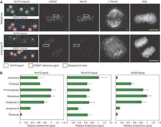
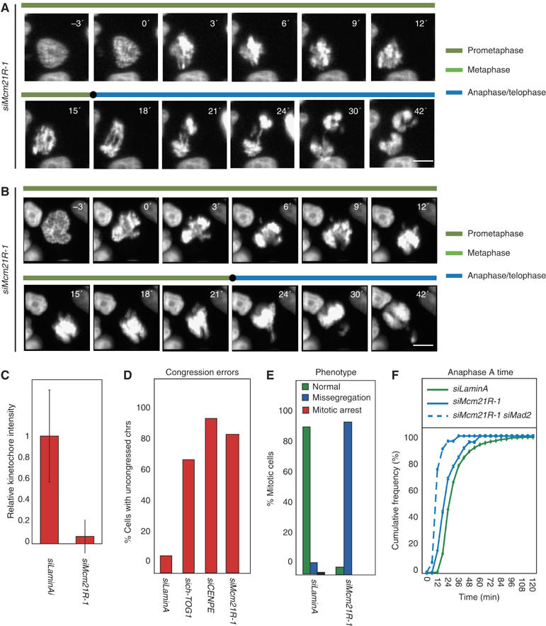
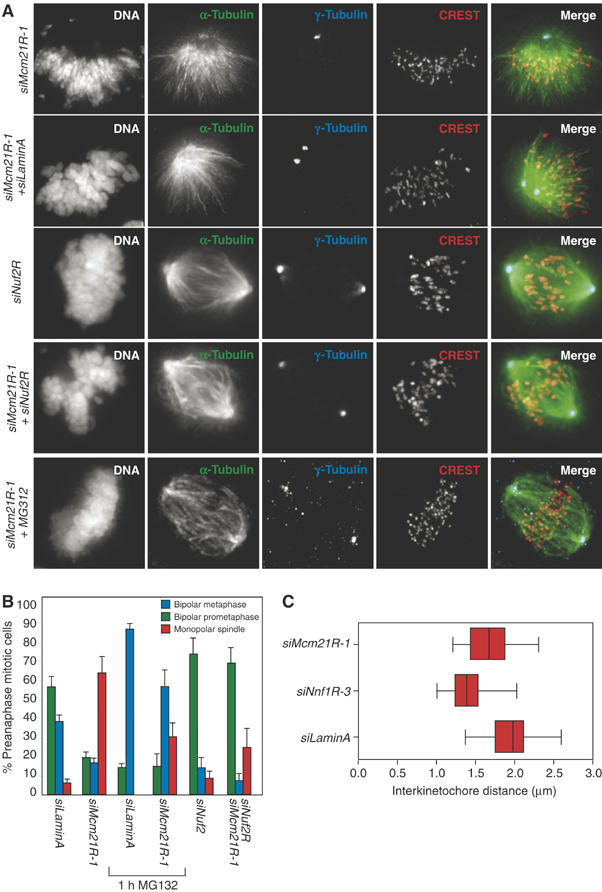
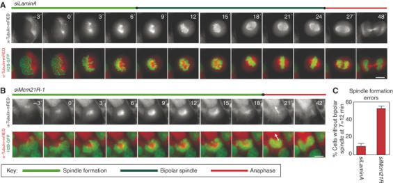
Kinetochores (KTs) assemble on centromeric DNA, bi-orient paired sister chromatids on spindle microtubules (MTs) and control cell-cycle progression via the spindle assembly checkpoint. Genetic and biochemical studies in budding yeast have established that three 'linker' complexes, MIND, COMA and NDC80, play essential but distinct roles in KT assembly and chromosome segregation. To determine whether similar linker activities are present at human KTs, we have compared the functions of Nnf1R and Mcm21R, recently identified MIND and COMA subunits, and Nuf2R, a well-characterized NDC80 subunit. We find that the three proteins bind to KTs independent of each other and with distinct cell-cycle profiles. MT-KT attachment is aberrant in Nnf1R- and Mcm21R-depleted cells, whereas it is lost in the absence of Nuf2R. Defective attachments in Nnf1R-depleted cells prevent chromosome congression, whereas those in Mcm21R-depleted cells interfere with spindle assembly. All three human KT proteins are necessary for correct binding of spindle checkpoint proteins to KTs. The differing functions and KT-binding properties of Nnf1R, Mcm21R and Nuf2R suggest that, like their yeast counterparts, the proteins act independent of each other in KT assembly, but that their combined activities are required for checkpoint signaling.


Figures

Figure 1
Figure 1
Localization of Nnf1R and Mcm21R in mitosis. (A) Immunoblots of lysates from cells transfected with siLaminA, siNnf1R-3 or siMcm21R-1 and probed with antibodies as indicated. (B) HeLa cells were stained with DAPI, γ-tubulin for spindle poles (blue), CREST antisera for KTs (red) and Nnf1R or Mcm21R antisera (green). As a control, localization of Nnf1R and Mcm21R was determined following MTs depolymerization with nocodazole for 1 h. (scale for insets). (C) Immunofluorescence images of prometaphase cells following transfection with siLaminA (negative control), siNnf1R-3 or siMcm21R-1 and stained with CREST (red) and Nnf1R or Mcm21R antisera (green) as indicated. Scale bars=10 μm.
Figure 2
Figure 2
Cell-cycle localization of Nnf1R, Mcm21R and Nuf2R during the mitotic cell cycle. (A) Levels of Nnf1R, Nuf2R and Mcm21R on KTs were determined from deconvolved 3D reconstructions of cells individually stained with DAPI, CREST antisera (KTs) and Nnf1R, Nuf2R or Mcm21R antisera. The intensity of KT signal (green) was determined relative to a CREST reference (red) after background correction (blue) (see Supplementary data for details). Box: 0.5 × 0.5 μm. Scale bar=10 μm. (B) Quantification of Nnf1R, Mcm21R and Nuf2R protein levels at KTs during different cell-cycle stages. Error bars show s.e.m. from repeat determinations in multiple cells.
Figure 3
Figure 3
Chromosome misalignment and missegregation in cells depleted of Nnf1R. (A) Successive frames every 3 or 4 min from live-cell movies of H2B-GFP-expressing HeLa cells transfected with either siLaminA, siNnf1R-1 or siNnf1R-3. Scale bar indicates 10 μm. (B) Cumulative frequency plots of anaphase onset times in siRNA-treated cells, as indicated, with NBD=T0, as determined from live-cell movies. (C) Fraction of cells, treated with siRNAs as indicated, having uncongressed chromosomes at T=24 min. (D) Mitotic phenotype following treatment with siRNAs as indicated. Cells were scored as either having undergone normal anaphase (green), having missegregated chromosomes in anaphase (blue) or becoming arrested prior to anaphase onset (as scored at T=120 min). (E) Quantification of Nnf1R protein levels, as described in Figure 2A, on KTs on prometaphase in siLaminA-, siNnf1R-1- or siNnf1R-3-treated cells.
Figure 4
Figure 4
Spindle checkpoint signaling fails following depletion of Nnf1R. (A, C) Cumulative frequency plots of anaphase onset times in siRNA-treated cells, as indicated, with NEB=T0, as determined from live-cell movies. (B) Percentage mitotic arrest in siLaminA-, siNnf1R-1-, siNnf1R-3 or siMad2-transfected cells treated with nocodazole for 16 h. (D) siLaminA-, siNnf1R-1- or siNnf1R-3-treated cells stained with CENP-A (KT marker), Mad2, Mad1, Bub1 or BubR1 antisera following treatment with nocodazole and MG132. Scale bar=10 μm. (E) Quantification of immunofluorescence signals as described in Figure 2A.
Figure 5
Figure 5
Nnf1R depletion leads to defects in KT-based force generation. (A) Chromosomes in metaphase cells were counted as unaligned if they were located outside of the central 30% of the mitotic spindle (dotted lines in schematic) or if their KTs were aligned perpendicular to the spindle axis. (B) Fraction of cells, treated with siLaminA, siNnf1R-1 or siNnf1R-3, having uncongressed chromosomes following 1 h treatment with MG132. (C) Representative immunofluorescence images of cells treated as described in (C) and stained with DAPI (blue), β-tubulin (green) and CENP-E (red) antisera. Scale bar=10 μm. (D, E) Inter-KT distances of chromosomes from cells arrested at metaphase with MG132 and treated with nocodazole or, siLaminA or siNnf1R-3. Inter-KT distances were measured using the outer KT marker CENP-E. They are shown as whisker plots and were derived by measuring at least 40 KT pairs in a total of eight cells. The central bar indicates the mean, the red box the 50th central percentile and the error bars the extremes.
Figure 6
Figure 6
Mcm21R depletion causes chromosome misalignment, missegregation and bipolar spindle assembly failure. (A, B) Successive frames every 3 min from live-cell movies of H2B-GFP-expressing HeLa cells transfected with siMcm21R-1. Scale bar=10 μm. (C) Quantification of Mcm21R protein levels, as described in Figure 2A, on KTs of prometaphase cells in siLaminA- or siMcm21R-1 treated cells. (D) Cumulative frequency plots of anaphase onset times in siRNA-treated cells, as indicated, with NBD=T0, as determined from live-cell movies. Note that siMcm21R-1 does not affect mitotic timing as opposed to siMad2 (solid blue lines). (E) Fraction of cells, treated with siRNA as indicated having uncongressed chromosomes at T=24 min. (F) Mitotic phenotype following treatment with siRNA as indicated. Cells were scored as either having undergone normal anaphase (green), having missegregated chromosomes in anaphase (blue) or becoming arrested prior to anaphase onset (as scored at T=120 min).
Figure 7
Figure 7
Spindle checkpoint failure in Mcm21R-depleted cells. (A) Percentage mitotic arrest in siLaminA-, siMcm21R-1- or siMad2-transfected cells treated with nocodazole for 16 h. (B) Cumulative frequency plots of anaphase onset times in siRNA-treated cells, as indicated, with NBD=T0, as determined from live-cell movies. (C) Quantification, as described in Figure 2A, of Bub1, BubR1, Mad1 and Mad2 proteins levels on KTs following siLaminA or siMcm21R-1 treatment. (D) Representative immunofluorescence images of cells from (C) stained with CREST, Mad2, Mad1, Bub1 or BubR1 antisera following treatment with nocodazole and MG132. Scale bar=10 μm.
Figure 8
Figure 8
Monopolar spindles in Mcm21R-depleted cells are suppressed by loss of MT–KT attachment. (A) Typical siMcm21R-1, siLaminA/siNuf2R, siLaminA or siMcm21R-1/siNuf2R metaphase cells stained with antisera against γ-tubulin or CREST (red), β-tubulin (green) and DAPI. siMcm21R-1/siLaminA cells were also stained for immunofluorescence following treatment with MG132 for 1 h (see sixth row). Scale bar indicates 10 μm. (B) Fraction of cells with either normal metaphase spindles (blue bars), bipolar prometaphase spindles (green bars) or monopolar spindles (red bars) determined following treatment with siRNAs as indicated in the presence and absence of MG132. (C) Inter-KT distances were calculated as described in Figure 4A following treatment with siLaminA, siNnf1R-3 or siMcm21R-1 after incubation with MG132 for 1 h.
Figure 9
Figure 9
Dynamics of spindle assembly in Mcm21R-depleted cells. Successive frames every 3 min from live-cell movies of H2B-GFP/α-tubulin-mRED-expressing HeLa cells transfected with siLaminA (A) or siMcm21R-1 (B). Shown are the composite images for H2B-GFP (green) and α-tubulin-mRED (red) or the single channel for α-tubulin-mRED (lower panels). Arrows indicate anaphase movement in siMcm21R-1-treated cells. Note chromosome decondensation in both cells 18 min after anaphase onset. Scale bar=10 μm. (C) Quantification of bipolar spindle formation errors at T=12 min after NBD.
Figure 10
Figure 10
Independent recruitment of Mcm21R, Nnf1R and Nuf2R onto KTs. Representative images of cells treated with siLaminA (A), siNnf1R-3 (B), siNuf2R (C) or siMcm21R-1 (D) and stained with Nnf1R, Mcm21R or Nuf2R antisera. Scale bar=10 μm. (EG) Quantification of KT signals for Nnf1R, Mcm21R and Nuf2R as described in Figure 2A–D.
Figure 11
Figure 11
Speculative model for the interface between spindle checkpoint and core KT factors. (A) Model of vertebrate KT organization based on electron micrographs and adapted from (Emanuele et al, 2005). (B) Assembly model for core KT components and spindle checkpoint components. Black arrows represent dependencies; a red ‘X' indicated a lack of dependency (Martin-Lluesma et al, 2002; Liu et al, 2003; Meraldi et al, 2004; Kops et al, 2005). (C) Model for the effect of partial and complete KT protein depletion on the spindle checkpoint.

References

    1. Bucciarelli E, Giansanti MG, Bonaccorsi S, Gatti M (2003) Spindle assembly and cytokinesis in the absence of chromosomes during Drosophila male meiosis. J Cell Biol 160: 993–999 - PMC - PubMed
    1. Buffin E, Lefebvre C, Huang J, Gagou ME, Karess RE (2005) Recruitment of Mad2 to the kinetochore requires the Rod/Zw10 complex. Curr Biol 15: 856–861 - PubMed
    1. Chan GK, Liu ST, Yen TJ (2005) Kinetochore structure and function. Trends Cell Biol 15: 589–598 - PubMed
    1. Cheeseman IM, Niessen S, Anderson S, Hyndman F, Yates JR III, Oegema K, Desai A (2004) A conserved protein network controls assembly of the outer kinetochore and its ability to sustain tension. Genes Dev 18: 2255–2268 - PMC - PubMed
    1. Ciferri C, De Luca J, Monzani S, Ferrari KJ, Ristic D, Wyman C, Stark H, Kilmartin J, Salmon ED, Musacchio A (2005) Architecture of the human ndc80-hec1 complex, a critical constituent of the outer kinetochore. J Biol Chem 280: 29088–29095 - PubMed

Publication types

MeSH terms