. 2006 Sep 6;25(17):4131-41.
doi: 10.1038/sj.emboj.7601272. Epub 2006 Aug 24.

Epididymal expression of the forkhead transcription factor Foxi1 is required for male fertility

Affiliations

Epididymal expression of the forkhead transcription factor Foxi1 is required for male fertility

Sandra Rodrigo Blomqvist et al. EMBO J. .

Abstract

An essential aspect of male reproductive capacity is the immediate availability of fertilization-ready spermatozoa. To ensure this, most mammals rely on post-testicular sperm maturation. In epididymis, germ cells are matured and stored in a quiescent state that readily can be altered to produce active spermatozoa. This depends on active proton secretion into the epididymal lumen. We have identified Foxi1 as an important regulator of gene expression in narrow and clear cells-the major proton secretory cells of epididymal epithelia. Foxi1 appears to be required for the expression of the B1-subunit of the vacuolar H+ -ATPase proton pump and for carbonic anhydrase II as well as the chloride/bicarbonate transporter pendrin. Using transfection experiments, we have identified a Foxi1 binding cis-element in the ATP6V1B1 (encoding the B1-subunit) promoter that is critical for reporter gene activation. When this site is mutated to eliminate Foxi1 binding, activation is also abolished. As a consequence of defect Foxi1-dependent epididymal sperm maturation, we demonstrate that spermatozoa from Foxi1 null males fail to reach the female genital tract in sufficient number to allow fertilization.


Figures

Figure 1
Figure 1
Foxi1 mRNA and protein expression in epididymis. Line drawing showing epididymal regions (A). Northern blot analysis identifies Foxi1 mRNA in epididymis but not in testis (B). Foxi1 cRNA in situ hybridization on wt epididymal tissue sections identified scattered Foxi1 positive cells (dark blue in C–E) while tissue sections from Foxi1−/− epididymis lacked Foxi1 (FH). Immunohistochemistry using antiserum against Foxi1 (green) showed Foxi1 protein in scattered cells in initial segment (I), caput (J), corpus (K) and cauda (L). No Foxi1 protein could be found in tissue sections derived from Foxi1−/− males (MP). The nuclei (blue) were visualized using To-Pro 3. Scale bars: 10 μm (L: lumen).
Figure 2
Figure 2
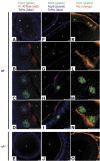
Confocal analysis of Foxi1, H+-ATPase, Aqp9 and Pds expression in wt and Foxi1−/− epididymides. In confocal images of wt epididymal sections, Foxi1 (green) protein was exclusively found in H+-ATPase (red) immunoreactive cells (AD). Foxi1 is localized to the nuclei, which are stained blue (To-Pro 3), while the proton pump is found on the apical/luminal side of the cell. Neither Foxi1 nor H+-ATPase is expressed in Foxi1−/− epididymis (E). aqp9 (purple) is expressed in the brush border membrane (BBM) of principal cells in epididymides derived from both wt (FI) and Foxi1−/− mice (J). These cells are negative for Foxi1. Cells without Aqp9 staining in their BBM have Foxi1 (green) protein in their nuclei (F–I). Pendrin (Pds; orange) is found both in the subepithelial sepulae of connective tissue and in Foxi1 (green) positive epithelial cells (KN). In Foxi1−/− epididymis the epithelial expression of Pds (orange) is lost (O). While Aqp9 (purple) is found in the BBM of principal cells in both Foxi1 wt and −/− epididymis epithelial H+-ATPase and Pds proteins are exclusively expressed in Foxi1 positive cells in wt epididymis and lost in Foxi1 deficient epididymis. Scale bars: 10 μm (L: lumen).
Figure 3
Figure 3
Identification of Foxi1 and Pds expressing cells. In situ hybridization using a Foxi1 cRNA probe (black) identifies a cell population in caput epithelia that transcribes Foxi1 mRNA (A). (BD) Confocal images using the same sections as in (A) for immunohistochemistry with H+-ATPase and Pds specific ab on slides previously exposed to Foxi1 cRNA in situ hybridization. We locate H+-ATPase (red, B, D) and Pds (green, C, D) to cells that are the same as the Foxi1 postitive cells in (A) (black, A). Thus, epithelial Pds, H+-ATPase and Foxi1 are all expressed in epididymal narrow and clear cells. Pds (green) staining is also seen in subepithelial connective tissue (C, D). In upper right corners of (B–D) are higher magnifications of representative clear cells. Scale bars: 10 μm (L: lumen).
Figure 4
Figure 4
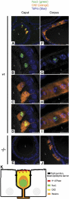
Distribution of CAII positive cells in wt and Foxi1−/− epididymis. In caput, CAII (orange) is expressed in the cytoplasm of cells that are immunoreactive for Foxi1 (green) (AD). Some CAII staining is also seen in nonepithelial cells. These cells are identified as erythrocytes, since they lack nucleus (stained blue using To-Pro 3), CAII is known to be highly expressed in the red blood cells. In corpus CAII (orange) is found in both Foxi1 (green) positive and negative cells (FI). In epididymes derived from Foxi1 deficient males no CAII (orange) protein is found in the caput epithelium (E) while CAII positive cells are present in Foxi1−/− corpus (J) and cauda (not shown). Scale bars: 10 μm (L: lumen). A schematic view of the proton secreting cell in epididymis (K; narrow and clear cells). Tight junctions constituting the blood–epididymis barrier (black rectangles) and approximate location of the Foxi1 dependent proteins are shown.
Figure 5
Figure 5
Foxi1 expression precedes that of Pds, CAII and H+-ATPase. In real-time RT–PCR quantification assays (TaqMan) of RNA from genital ridges and epididymes at time points from E12.5 to adult, measurable levels of Foxi1 transcripts are first detected at P5 and increase during postnatal development (A). This is in accordance with immunohistochemistry results (BM). At P5, only Foxi1 (green) is found in a few cells of wt epididymis epithelium (B–D). Extra-epithelial Pds (purple) derived form connective tissue is seen at all time-points regardless of genotype (B, E, F, I, J, M). Foxi1 deficient P5 epididymis lacks epithelial Pds (purple; E). At P10, Pds (purple) is found in epithelial cells, colocalized with Foxi1 (green) positive cells (F). CAII (orange) protein is also expressed at P10, in the cytoplasm of Foxi1 (green) expressing cells (G). Also a higher number of wt epididymis cells are Foxi1 (green) positive (H). At P15, Pds (purple, J) and CAII (orange, K) are present in Foxi1 (green) immune-reactive cells. At this time-point H+-ATPase (red) is identified, located in the luminal part of the Foxi1 (green) expressing cells (L). No epithelial expression of Foxi1 (E, I, M, left panel), Pds (E, I, M, right panel), CAII (E, I, M, left panel) and H+-ATPase (E, I, M, left panel) could be found on Foxi1−/− tissue sections at any of these time points. Nuclei are stained blue. **P<0.01. Scale bar: 10 μm (L: lumen).
Figure 6
Figure 6
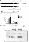
FOXI1 specifically binds to and activates the ATP6V1B1 promoter. Schematic view of the reporter gene constructs used in the luciferase assays (A). A 1.0 kb nonmutated luciferase reporter construct (5-ATP6V1B1) and a mutated construct (5ATP6V1B1 TTT>GGG), in which the core T-triplet at position −102 to −96 has been replaced by GGG (A). Transient transfection using COS7 cells. When co-transfected with a FOXI1 expression vector (40, 80 and 100 ng) the wt reporter construct (5ATP6V1B1) showed a significant induction of reporter gene activity (***P<0.001). In contrast, the mutated variant (5ATP6V1B1 TTT>GGG) displayed no activity above what was generated with 100 ng of expression vector without insert. Reporter gene activity is shown as fold induction relative to activity generated in transfections using 100 ng of expression vector void of FOXI1 (B). EMSA of in vitro transcribed/translated FOXI1 protein (see Materials and methods) using radioactively labeled oligonucleotides encoding the putative FOXI1 binding site (−102 to −96) of the 5ATP6V1B1 promoter as well as a mutated site (same as in A, B). As can be deduced wt labeled oligonucleotide together with FOXI1 protein generates a band with altered mobility (C; lane 2). Using increasing amounts of wt nonlabeled (cold) oligonucleotide as competitor (lanes 3–6; 2-, 10-, 20- and 50-fold molar excess) demonstrates a gradually decreased intensity of the signal as would be expected for a specific DNA–protein interaction. In a similar experiment using mutated oligonucleotide instead of wt (lanes 7–10), no specific competition could be detected.
Figure 7
Figure 7
Wt and Foxi1−/− sperm morphology and quantification. Light microscopy studies of sperms from wt (A, B) and Foxi1 deficient (C, D) males revealed a significantly higher number of sperms with tail angulation in −/− mice (C–E). Direct sperm counts from flushed uteruses (F) revealed a significant difference (*P<0.05) in number of sperms derived from wt females mated with Foxi1−/− males as compared with wt males. As a likely sign of altered luminal microenvironment Foxi1−/− males have increased epididymis/body weight ratio (G; **P<0.01) as well as an increased epididymal area (H). Scale bar: 30 μm (A, C), 20 μm (B, D).

References

    1. Acott TS, Carr DW (1984) Inhibition of bovine spermatozoa by caudal epididymal fluid: II. Interaction of pH and a quiescence factor. Biol Reprod 30: 926–935 - PubMed
    1. Al-Awqati Q (1996) Plasticity in epithelial polarity of renal intercalated cells: targeting of the H(+)-ATPase and band 3. Am J Physiol 270: C1571–C1580 - PubMed
    1. Bedford JM (1975) Maturation, transport, and fate of spermatozoa in the epididymis. In Handbook of Physiology Section 7: Endocrinolology, Vol. V. Male Reproductive System, Greep RO, Astwood EB (eds). pp 303–317. Washington DC: American Physiology Society
    1. Blomqvist SR, Vidarsson H, Fitzgerald S, Johansson BR, Ollerstam A, Brown R, Persson AE, Bergstrom GG, Enerback S (2004) Distal renal tubular acidosis in mice that lack the forkhead transcription factor Foxi1. J Clin Invest 113: 1560–1570 - PMC - PubMed
    1. Breton S, Smith PJ, Lui B, Brown D (1996) Acidification of the male reproductive tract by a proton pumping (H+)-ATPase. Nat Med 2: 470–472 - PubMed

Publication types

MeSH terms