. 2006 Sep;116(9):2543-51.
doi: 10.1172/JCI26936. Epub 2006 Aug 24.

Relating TCR-peptide-MHC affinity to immunogenicity for the design of tumor vaccines

Affiliations

Relating TCR-peptide-MHC affinity to immunogenicity for the design of tumor vaccines

Rachel H McMahan et al. J Clin Invest. 2006 Sep.

Abstract

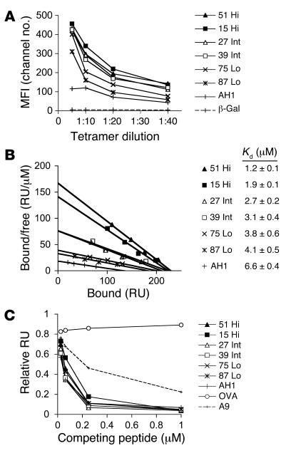
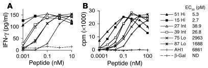
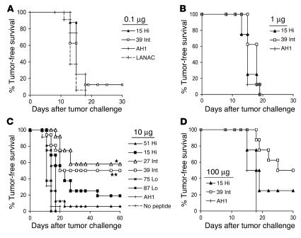
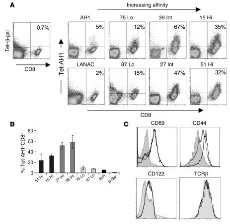
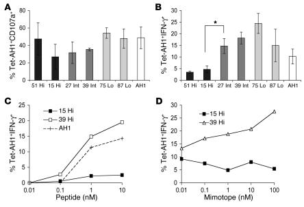
One approach to enhancing the T cell response to tumors is vaccination with mimotopes, mimics of tumor epitopes. While mimotopes can stimulate proliferation of T cells that recognize tumor-associated antigens (TAAs), this expansion does not always correlate with control of tumor growth. We hypothesized that vaccination with mimotopes of optimal affinity in this interaction will improve antitumor immunity. Using a combinatorial peptide library and a cytotoxic T lymphocyte clone that recognizes a TAA, we identified a panel of mimotopes that, when complexed with MHC, bound the TAA-specific TCR with a range of affinities. As expected, in vitro assays showed that the affinity of the TCR-peptide-MHC (TCR-pMHC) interaction correlated with activity of the T cell clone. However, only vaccination with mimotopes in the intermediate-affinity range elicited functional T cells and provided protection against tumor growth in vivo. Vaccination with mimotopes with the highest-affinity TCR-pMHC interactions elicited TAA-specific T cells to the tumor, but did not control tumor growth at any of the peptide concentrations tested. Further analysis of these T cells showed functional defects in response to the TAA. Thus, stimulation of an antitumor response by mimotopes may be optimal with peptides that increase but do not maximize the affinity of the TCR-pMHC interaction.


Figures

Figure 1
Figure 1. Affinity characterization of mimotope peptides.
(A) Relative avidities of the TCR-pMHC interactions were determined by staining a T cell clone that recognizes the AH1 peptide with increasing concentrations of Ld-tet incubated with the indicated peptides. The H-2Ld–restricted β-gal peptide (64) did not bind the TCR from the T cell clone. Data are presented as mean fluorescence intensity (MFI) and are representative of 3 independent experiments. (B) Affinity binding analysis of the pMHC complexes for the TCR from the T cell clone were determined using SPR. Increasing concentrations of soluble H-2Ld monomers incubated with peptide were exposed to the immobilized TCR on a biosensor surface. Equilibrium-binding measurements were used to construct Scatchard plots. Kd values were calculated by nonlinear regression analysis of the response units plotted against H-2Ld concentration (Prism, version 4.0; GraphPad Software). (C) Relative affinities of the peptides for H-2Ld were determined using competition assays. The indicated peptide concentrations and 40 μM monomeric H-2Ld were exposed to the immobilized pMCMV-C4 peptide on a biosensor surface. The amount of H-2Ld bound to pMCMV-C4 is reflected by the change in RU after 1 minute. Data are presented as RU relative to binding of empty H-2Ld to the MCMV-C4 peptide. Hi, high affinity; Int, intermediate affinity; Lo, low affinity.
Figure 2
Figure 2. Mimotopes potently activate the T cell clone.
The T cell clone was cultured with increasing concentrations of the AH1 peptide, mimotopes, or the negative control β-gal peptide. (A) Production of IFN-γ, as determined by ELISA, from the T cell clone in response to incubation for 24 hours with the indicated peptide. (B) After 24 hours, 3H-thymidine was added to the cultures, and 3H-thymidine incorporation was measured at 48 hours. Data are representative of 2 experiments per peptide, and each data point was performed in triplicate and averaged. ND, no data (EC50 was too low to be determined).
Figure 3
Figure 3. Vaccination with mimotopes of intermediate affinity most effectively protects against tumor challenge.
BALB/c mice were primed on days –17 and –10 with (A) 0 or 0.1 μg, (B) 1 μg, (C) 10 μg, or (D) 100 μg peptides and LANAC (adjuvant) or LANAC alone followed by injection of 5 × 10⁴ CT26 tumor cells on day 0 (subcutaneous, back left flank). Tumor-free survival was monitored and plotted using the Kaplan-Meier method (Prism, version 4.0; GraphPad Software). Survival among groups was compared using the log-rank test (Prism, version 4.0; GraphPad Software). *P < 0.004, **P < 0.006 versus mimotope 15. All vaccination groups consisted of 8 mice per peptide with the exception of the 10 μg dose, for which 16 mice per peptide were challenged (except for the mimotope 27, 75, and 87 groups, which each consisted of 12 mice).
Figure 4
Figure 4. The AH1-specific T cell response was strongest after priming with the intermediate-binding mimotopes.
(A) BALB/c mice with day 4 established tumors were injected with LANAC and the indicated peptides. On day 14, TILs were isolated and stained ex vivo with CD8 antibody and Ld-tet bound to either the AH1 or β-gal peptide. The percentages of tetramer-positive CD8+ cells are indicated. (B) Using the methods from A, the mean ± SEM of AH1-specific CD8+ TILs was calculated for 4 mice per vaccine. (C) Ld-tet/AH1–positive CD8+ T cells from tumors of mice vaccinated with AH1 (filled), mimotope 39 (black line), or mimotope 15 (gray line) peptides were stained ex vivo with CD69, CD44, CD122, and TCRβ antibodies. Staining of activation markers was not performed for the other mimotopes.
Figure 5
Figure 5. TILs elicited by high-affinity mimotopes produce cytotoxic granules, but have impaired production of IFN-γ in response to antigen.
BALB/c mice with day 4 established CT26 tumors were primed with LANAC alone or LANAC with the indicated peptide on days 4 and 6. (A and B) CD8+ TILs were isolated on day 14 and stimulated with 1 μM AH1 peptide for 5 hours at 37°C. Cells were stained with CD8 antibody and Ld-tet/AH1 as well as (A) CD107a, a marker of cytotoxic granules, or (B) IFN-γ antibody. The mean ± SEM was calculated for 4 mice per peptide. *P = 0.0125. Functional analysis of TILs from mice vaccinated with LANAC alone was not possible due to low numbers of AH1-specific T cells. (C and D) CD8+ TILs isolated from mice vaccinated with AH1, mimotope 39, or mimotope 15 were stimulated with graded concentrations of (C) the AH1 peptide or (D) the mimotope used for vaccination and stained for intracellular IFN-γ after 5 hours at 37°C.

References

    1. Dunn G.P., Old L.J., Schreiber R.D. The three Es of cancer immunoediting. Annu. Rev. Immunol. 2004;22:329–360. - PubMed
    1. Marincola F.M., Jaffee E.M., Hicklin D.J., Ferrone S. Escape of human solid tumors from T-cell recognition: molecular mechanisms and functional significance. Adv. Immunol. 2000;74:181–273. - PubMed
    1. Gorelik L., Flavell R.A. Immune-mediated eradication of tumors through the blockade of transforming growth factor-beta signaling in T cells. Nat. Med. 2001;7:1118–1122. - PubMed
    1. Niethammer A.G., et al. A DNA vaccine against VEGF receptor 2 prevents effective angiogenesis and inhibits tumor growth. Nat. Med. 2002;8:1369–1375. - PubMed
    1. Novellino L., Castelli C., Parmiani G. A listing of human tumor antigens recognized by T cells: March 2004 update. Cancer Immunol. Immunother. 2005;54:187–207. - PMC - PubMed

Publication types