. 2006 Sep;116(9):2510-20.
doi: 10.1172/JCI29128. Epub 2006 Aug 24.

Casq2 deletion causes sarcoplasmic reticulum volume increase, premature Ca2+ release, and catecholaminergic polymorphic ventricular tachycardia

Affiliations

Casq2 deletion causes sarcoplasmic reticulum volume increase, premature Ca2+ release, and catecholaminergic polymorphic ventricular tachycardia

Björn C Knollmann et al. J Clin Invest. 2006 Sep.

Abstract

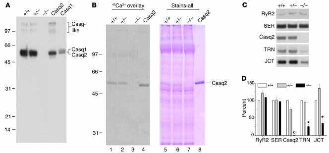
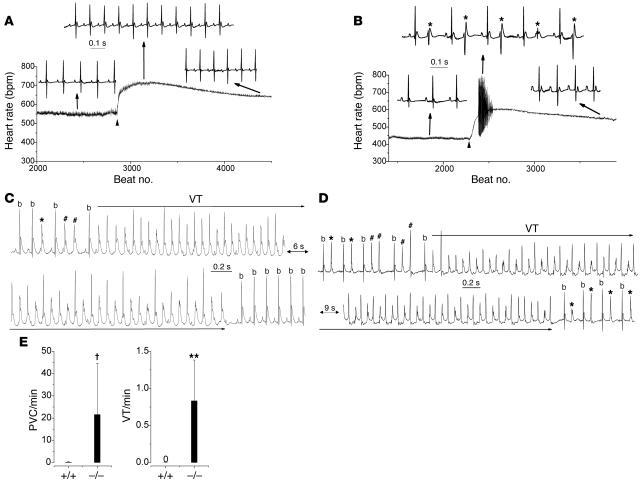
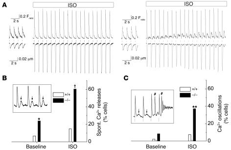
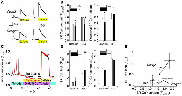
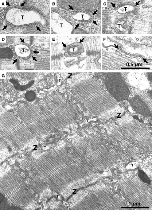
Cardiac calsequestrin (Casq2) is thought to be the key sarcoplasmic reticulum (SR) Ca2+ storage protein essential for SR Ca2+ release in mammalian heart. Human CASQ2 mutations are associated with catecholaminergic ventricular tachycardia. However, homozygous mutation carriers presumably lacking functional Casq2 display surprisingly normal cardiac contractility. Here we show that Casq2-null mice are viable and display normal SR Ca2+ release and contractile function under basal conditions. The mice exhibited striking increases in SR volume and near absence of the Casq2-binding proteins triadin-1 and junctin; upregulation of other Ca2+ -binding proteins was not apparent. Exposure to catecholamines in Casq2-null myocytes caused increased diastolic SR Ca2+ leak, resulting in premature spontaneous SR Ca2+ releases and triggered beats. In vivo, Casq2-null mice phenocopied the human arrhythmias. Thus, while the unique molecular and anatomic adaptive response to Casq2 deletion maintains functional SR Ca2+ storage, lack of Casq2 also causes increased diastolic SR Ca2+ leak, rendering Casq2-null mice susceptible to catecholaminergic ventricular arrhythmias.


Figures

Figure 1
Figure 1. Generating the Casq2 allele.
(A) The Casq2 exon 1 sequence as determined by 5′ RACE. Four transcriptional starts were identified and are marked with filled arrowheads. Nucleotide number 1 represents the 5′ end of the longest identified transcript. Exon 1 includes 3 in-frame ATG translational starts (underlined). Only the second ATG is conserved in other vertebrates, and only this ATG is predicted to encode a leader sequence that would appropriately target the nascent CASQ2 peptide to the SR. (B) Wild-type (i) and mutant (ii) alleles of the Casq2 locus are depicted. The Casq2 locus spans more than 60 kb and includes 11 exons (vertical bars). Exon 1 encodes the ATG initiation codon and the first 78 amino acids. The Casq2 allele is a 1.1-kb deletion that removes 561 bp of upstream sequences, including the presumptive Casq2 promoter (open oval) as well as the entire 431-bp exon 1 and 107 bp of intron 1.
Figure 2
Figure 2. Casq2–/– hearts lack calsequestrin, display no apparent upregulation of other SR Ca2+-binding proteins, and have decreased triadin 1 and junctin protein levels.
(A) Forty micrograms of homogenate protein from Casq2+/+, Casq2+/–, and Casq2–/– hearts and 30 μg of microsomal protein from control membranes from mouse heart (Casq2) and skeletal muscle (Casq1) were electrophoresed per lane and probed with anti-calsequestrin antibody. Cardiac (Casq2), skeletal muscle (Casq1), and Casq-like proteins are indicated. (B) 45Ca2+ overlay and Stains-all staining of SR membrane proteins obtained from Casq2+/+, Casq2+/–, and Casq2–/– hearts. Seventy-five micrograms of SR membrane protein was loaded per lane in duplicate and subjected to SDS-PAGE, then one-half of the gel was processed for 45Ca2+ overlay (left) and the other half stained with Stains-all (right). One microgram of purified canine Casq2 was also run as an internal standard. (C) Immunoblot detection of SR proteins in microsomes isolated from 10 Casq2+/+, 10 Casq2+/–, and 10 Casq2–/– hearts. Forty micrograms of microsomal protein were electrophoresed per lane, transferred to nitrocellulose paper, and probed with the antibodies indicated on the left. (D) Quantification of protein expression levels. Data represent average values for 4 hearts per genotype expressed relative to Casq2+/+ values. RyR2, cardiac isoform of the RyR; SER, SERCA2a or cardiac isoform of the Ca2+ pump; TRN, triadin 1 or major cardiac isoform of triadin; JCT, junctin; *P < 0.05.
Figure 3
Figure 3. Casq2–/– mice display catecholaminergic ventricular ectopy and exercise-induced polymorphic VT.
(A and B) Continuous heart rate plot and examples of surface ECG (lead 1) recordings from an anesthetized Casq2+/+ (A) and Casq2–/– mouse (B) injected with the β-adrenergic receptor agonist isoproterenol (1.5 mg/kg i.p.; arrowhead). Note the multifocal PVCs (*) at the peak of the heart rate response in the Casq2–/–mouse. (C) Example of a telemetric ECG recording from a conscious Casq2–/– mouse obtained shortly after a treadmill exercise tolerance test. PVCs, couplets (#), and runs of polymorphic VT were frequently observed in Casq2–/–mice. All episodes of polymorphic VT reverted spontaneously back to sinus rhythm (b, sinus beat; lower record). (D) Example of bidirectional VT recorded in another Casq2–/– mouse. Bidirectional VT was initiated after several bigemini and couplets and terminated into stable bigemini. (E) Average rate of PVCs and VT episodes during a 10-minute period of post-exercise telemetric ECG recordings. n = 5 mice per genotype; P < 0.05, **P < 0.01.
Figure 4
Figure 4. Casq2–/– myocytes display spontaneous Ca2+ releases and triggered beats but largely maintain normal contractility, SR Ca2+ release amplitudes, and SR Ca2+ content.
(A) Examples of [Ca2+]i transients (top traces) and cell shortening (bottom traces) recorded from fura-2/AM–loaded, field-stimulated myocytes (1 Hz). Application of 1 μmol/l isoproterenol (ISO) significantly increased Ca2+ transients and cell shortening in both myocytes. Note that shortly after ISO application was started, only the Casq2–/–myocyte displayed spontaneous Ca2+ releases and aftercontractions of increasing amplitude following each paced twitch (vertical lines). (B and C) Comparison of the incidence of spontaneous (Spont.) Ca2+ release events (B) and Ca2+ oscillations (C) at baseline and in the presence of ISO. Data represent the fraction (%) of myocytes that displayed at least 1 event during a 20-second recording period. Insets show representative examples of spontaneous Ca2+ after-releases (arrows in B, inset) and Ca2+ oscillations (C, inset) induced by spontaneous Ca2+ releases and triggered beats (# in C, inset). *P < 0.05, **P < 0.01, P < 0.001, Casq2–/– versus Casq2+/+ myocytes by Fisher’s exact test. Casq2+/+ myocytes: n = 45 (baseline) and 27 (ISO); Casq2–/–myocytes: n = 71 (baseline) and 43 (ISO).
Figure 5
Figure 5. Casq2–/– myocytes have largely preserved SR Ca2+ release and SR Ca2+ content under basal conditions, but isoproterenol application causes increased SR Ca2+ leak.
(A) Representative examples of rapid application of caffeine (10 mmol/l) to a Casq2+/+ (top) and a Casq2–/– myocyte (bottom). Myocytes were field stimulated at 1 Hz to maintain consistent SR Ca2+ load. Note the increased twitch transient and caffeine-induced transients in the presence of ISO (1 μmol/l; right). The height of the caffeine-induced Ca2+ transient was used as a measure of total SR Ca2+ content (30). Fractional SR Ca2+ release was calculated by dividing the height of the last twitch transient by the height of the caffeine transient. (B) Comparison of average SR Ca2+ content (left) and fractional SR Ca2+ release (right). *P < 0.05, **P < 0.01. Casq2+/+ myocytes: n = 41 (baseline) and 27 (ISO); Casq2–/–myocytes: n = 70 (baseline) and 37 (ISO). (C) Protocol used to measure SR Ca2+ leak as described in ref. 32. Plasma membrane Ca2+ flux is eliminated by removal of extracellular Na+ and Ca2+. The drop in steady-state [Ca2+]i (double arrow) represents a shift of Ca2+ from the cytosol to the SR when RyR2 channels are inhibited by tetracaine (1 mmol/l) and was used as a measure of SR Ca2+ leak. (D) Comparison of average SR Ca2+ leak (left) and SR Ca2+ content in the presence of tetracaine (right). Note that when SR Ca2+ leak was blocked by tetracaine, SR Ca2+ content was not significantly different between the 2 groups. **P < 0.01. Casq2+/+ myocytes: n = 32 (baseline) and 45 (ISO); Casq2–/–myocytes: n = 29 (baseline) and 42 (ISO). (E) SR Ca2+ leak in the presence of ISO plotted as a function of SR Ca2+ content. Note that the SR Ca2+ leak of Casq2–/– myocytes remained SR load dependent but was shifted to the left compared with that of Casq2+/+ myocytes.
Figure 6
Figure 6. jSR lacks its visible content, and overall SR volume is increased in Casq2–/– ventricular myocytes.
Electron micrographs of left-ventricular myocytes from Casq2+/+ (A and B) and Casq2–/– (CG) hearts. Flat jSR cisternae are closely apposed to T tubules (T). In Casq2+/+ myocytes, cisternae are quite uniform in width and filled by calsequestrin, which is periodically arranged in small clumps (A and B, between arrows). “Feet” (RyRs) are present in the junctional gap between the 2 membranes. In Casq2–/– myocytes, the jSR has variable width and is apparently empty (CF, between arrows). (G) Longitudinal SR of a Casq2–/– myocyte in a section that was cut tangentially to the myofibrils. The SR forms a network with large fenestrations covering the A bands and continuing across Z lines (Z). In proximity to the T tubules, the SR runs in a transverse orientation, accompanying the T tubule profiles. The SR network is quite abundant in Casq2–/– myocytes (see data in Table 2).

References

    1. Scott B.T., Simmerman H.K., Collins J.H., Nadal-Ginard B., Jones L.R. Complete amino acid sequence of canine cardiac calsequestrin deduced by cDNA cloning. J. Biol. Chem. 1988;263:8958–8964. - PubMed
    1. Yano K., Zarain-Herzberg A. Sarcoplasmic reticulum calsequestrins: structural and functional properties. Mol. Cell. Biochem. 1994;135:61–70. - PubMed
    1. Cala S.E., Jones L.R. Rapid purification of calsequestrin from cardiac and skeletal muscle sarcoplasmic reticulum vesicles by Ca2+-dependent elution from phenyl-sepharose. J. Biol. Chem. 1983;258:11932–11936. - PubMed
    1. Campbell K.P., MacLennan D.H., Jorgensen A.O., Mintzer M.C. Purification and characterization of calsequestrin from canine cardiac sarcoplasmic reticulum and identification of the 53,000 dalton glycoprotein. J. Biol. Chem. 1983;258:1197–1204. - PubMed
    1. Jones L.R., Cala S.E. Biochemical evidence for functional heterogeneity of cardiac sarcoplasmic reticulum vesicles. J. Biol. Chem. 1981;256:11809–11818. - PubMed

Publication types

MeSH terms